Home
Images
Vectors
Videos
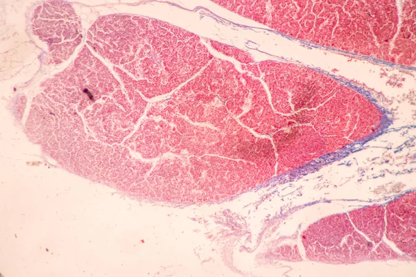
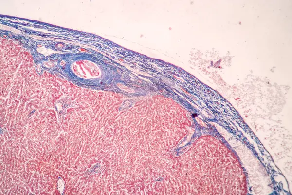
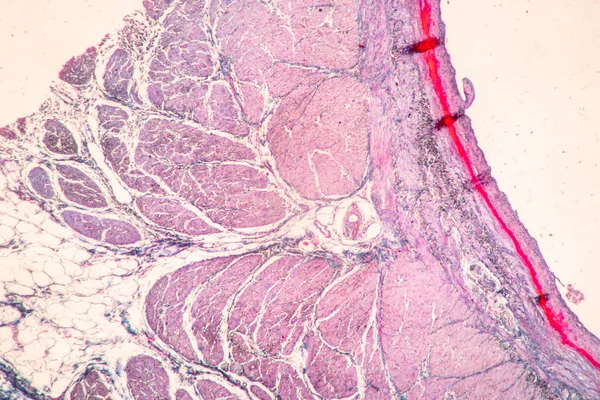
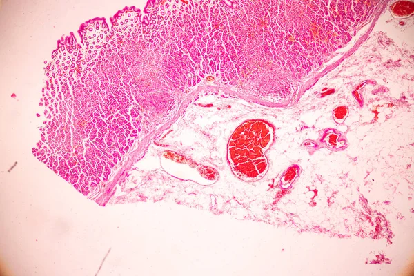
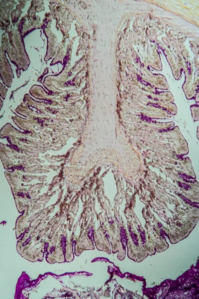
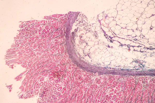
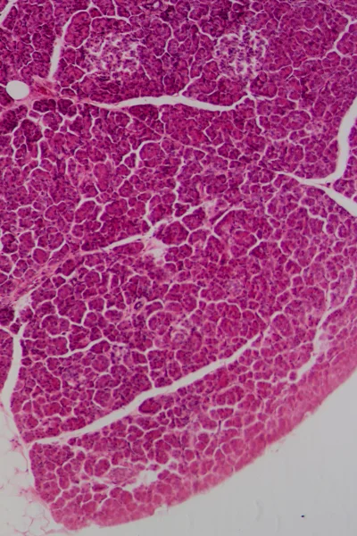
Stock image Pathology and Histology Tissue of Mammals under microscope.
Published:
Aug.05, 2024 09:44:38
Author:
p.thongdumhyu
Views:
0
Downloads:
0
File type:
image
/
jpg
File size:
18.66 MB
Orginal size:
6000
x
4000
px
Available sizes:
S
(500 x 333)
M
(1000 x 667)
L
(2000 x 1333)
XL
(6000 x 4000)
EL0
(6000 x 4000)
Level:
beginner
Download Image
Image topics:
Animal
Pattern
Mammal
Skin
Structure
Cross
Cell
Disease
Education
Sample
Biology
Laboratory
Science
Section
Study
Fiber
Organ
Tissue
Magnification
Anatomy
Surgery
Scientist
Microscope
Connective
Liver
Scalp
Types
Morphology
Hepatitis
Dermis
Pathology
Nerve
Intestine
Tumour
Colon
Photomicrograph
Collagen
Prostate
Histology
Squamous
Epithelium
Squamous Cell
Connective Tissue
Eosin
Hematoxylin
Hair Follicles
Myocytes
Squamous Cell Carcinoma
Elastic Cartilage
Similar stock images
Backgrounds Of Characteristics Tissue Of Stomach Human, Small Intestine Human, Pancreas Human And Large Intestine Human Under The Microscope In Lab.
6720 × 4480
Structure Of Tissue Of Spleen Human, Liver Human And Kidney Human Under The Microscope In Lab.
6720 × 4480
Backgrounds Of Characteristics Tissue Of Stomach Human, Small Intestine Human, Pancreas Human And Large Intestine Human Under The Microscope In Lab.
6720 × 4480
Tissue Of Stomach Human Under The Microscope In Lab.
4254 × 2836
Cross-section Through The Intestine With Glands 100x
3744 × 5616
Backgrounds Of Characteristics Tissue Of Stomach Human, Small Intestine Human, Pancreas Human And Large Intestine Human Under The Microscope In Lab.
6720 × 4480
Microscopic Section Of Liver Tissue
3168 × 4752
Section Of A Common Liver Fluke (Fasciola) Under The Microscope
4500 × 3000