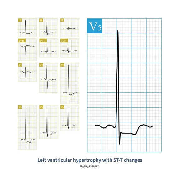
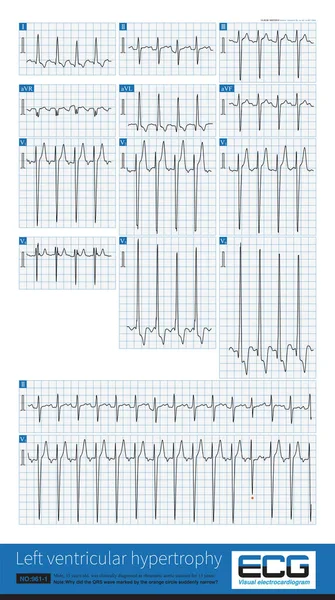
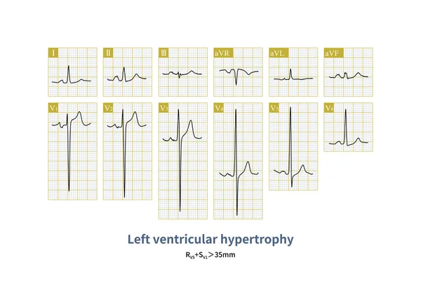
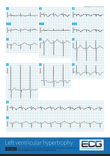
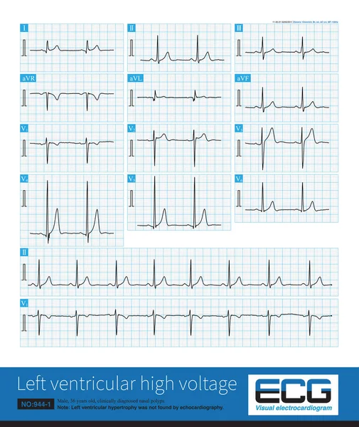
Stock image The ECG of a patient with patent ductus arteriosus indicates left ventricular hypertrophy without ST changing.
Published: Jun.07, 2023 06:13:01
Author: asia11m
Views: 9
Downloads: 0
File type: image / jpg
File size: 21.69 MB
Orginal size: 8000 x 15812 px
Available sizes:
Level: beginner