photo free
Home Images Vectors Videos

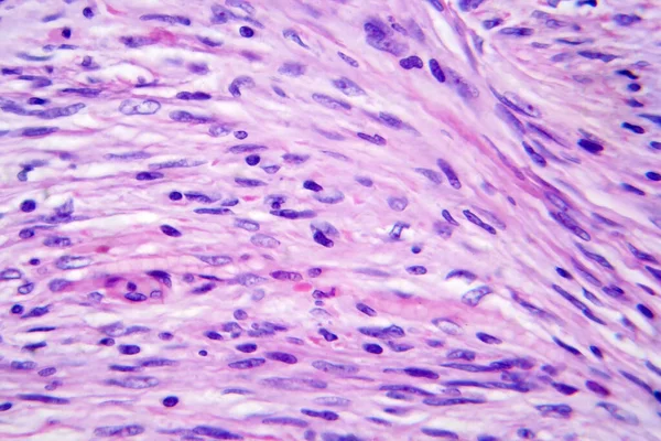
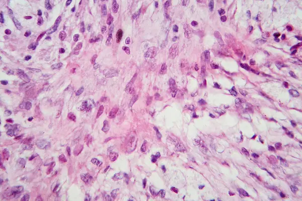
Stock image This photo shows the pink mucinous matrix and linear arrangement of mucinous tumor cells in atrial myxoma.Magnify 1000x.

Stock image This photo shows the pink mucinous matrix and linear arrangement of mucinous tumor cells in atrial myxoma.Magnify 1000x.
Published: May.30, 2024 13:31:06
Author: asia11m
Views: 0
Downloads: 0
File type: image / jpg
File size: 42.79 MB
Orginal size: 6000 x 7851 px
Available sizes:
S
(382 x 500)
M
(764 x 1000)
L
(1528 x 2000)
XL
(6000 x 7851)
EL0
(6000 x 7851)
Level: beginner
Download Image
Image topics:

Similar stock images

stock image This pathologic  photograph highlights the atrium Myxoma cell nest. Atrial myxoma is a benign tumor of the heart.

This Pathologic Photograph Highlights The Atrium Myxoma Cell Nest. Atrial Myxoma Is A Benign Tumor Of The Heart.

5500 × 7384
stock image This is a pathological photo of a human gout nodule, showing the formation of pink amorphous eosinophilic substances by urate crystals.Magnify 40x.

This Is A Pathological Photo Of A Human Gout Nodule, Showing The Formation Of Pink Amorphous Eosinophilic Substances By Urate Crystals.Magnify 40x.

6000 × 6000
stock image Meningioma, a slow-growing tumor that forms from the meninges, light micrograph, photo under microscope

Meningioma, A Slow-growing Tumor That Forms From The Meninges, Light Micrograph, Photo Under Microscope

4364 × 2909
stock image Photomicrograph showing histological features of benign prostatic hyperplasia. Enlarged prostate gland with nodular proliferation of glandular and stromal components.

Photomicrograph Showing Histological Features Of Benign Prostatic Hyperplasia. Enlarged Prostate Gland With Nodular Proliferation Of Glandular And Stromal Components.

7178 × 4786
stock image Photomicrograph of a giant cell tumor of bone, revealing numerous giant cells within a background of mononuclear cells and hemosiderin deposition.

Photomicrograph Of A Giant Cell Tumor Of Bone, Revealing Numerous Giant Cells Within A Background Of Mononuclear Cells And Hemosiderin Deposition.

7124 × 4749
stock image Photomicrograph of hepatic cavernous hemangioma, depicting dilated blood vessels in the liver tissue, characteristic of a benign tumor.

Photomicrograph Of Hepatic Cavernous Hemangioma, Depicting Dilated Blood Vessels In The Liver Tissue, Characteristic Of A Benign Tumor.

7124 × 4749
stock image Fibrous thyroiditis, light micrograph, photo under microscope. A disease characterized by chronic inflammation and fibrosis of the thyroid gland

Fibrous Thyroiditis, Light Micrograph, Photo Under Microscope. A Disease Characterized By Chronic Inflammation And Fibrosis Of The Thyroid Gland

4364 × 2909