Home
Images
Vectors
Videos
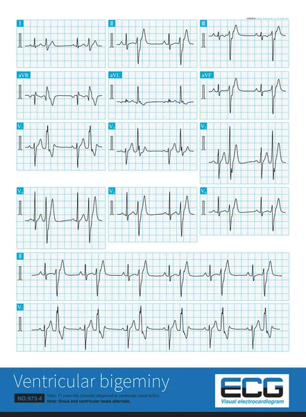
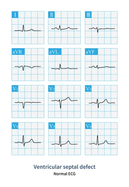
Stock image When a small ventricular septal defect has little or even no impact on hemodynamics, the patient's electrocardiogram can be completely or roughly normal.
Published:
Nov.08, 2023 09:02:03
Author:
asia11m
Views:
2
Downloads:
0
File type:
image
/
jpg
File size:
18.49 MB
Orginal size:
9000
x
12961
px
Available sizes:
S
(347 x 500)
M
(694 x 1000)
L
(1389 x 2000)
XL
(9000 x 12961)
EL0
(9000 x 12961)
Level:
beginner
Download Image
Image topics:
Blue
Graphic
Human
Health
Medicine
Medical
Cardiology
Hospital
Body
Disease
Education
Science
Death
Graph
Beat
Rhythm
Diagnosis
Heartbeat
Cardiogram
Cardiac
Ekg
Electrocardiogram
Ecg
Heart Disease
Heart Failure
Cardiac Arrest
Blue Line
Congenital Heart Disease
Left Ventricle
Right Ventricle
Dyspnea
Pulmonary Hypertension
Left Atrium
Cardiac Surgery
Sudden Death
Pulmonary Artery
Interventricular Septum
Right Atrium
Ventricular Septal Defect
ECG Monitoring
Left Ventricular Hypertrophy
Right Ventricular Hypertrophy
St Segment
Ecg Education
Qrs Wave
Left Ventricular Enlargement
Organic Heart Disease
Interventional Treatment
Similar stock images
A Patient With Extensive Anterior Myocardial Infarction Had Wide QRS Wave Tachycardia, That Is, Persistent Ventricular Tachycardia, With Atrioventricular Separation.
10000 × 8171
The Typical ECG For Left Ventricular Hypertrophy Is Left Ventricular High Voltage, Sometimes With Significant ST-T Changes, U-wave Changes, And QT Interval Prolongation.
8000 × 11283
Simple Left Ventricular High Voltage Is Only An ECG Phenomenon. The Subject Has No Pathological Left Ventricular Hypertrophy And No Other Abnormal ECG Changes.
10000 × 11864
A 17 Year Old Male Was Clinically Diagnosed As Ventricular Septal Defect And His ECG Was Ventricular Bigeminy.Ventricular Premature Contraction Originated In The Left Posterior Fascicular Region.
10000 × 13607
Sometimes, Because The QRS Axis Is In The Upper Left Quadrant, The High-amplitude R Wave Of Left Ventricular Hypertrophy Occurs In The Limb Leads, And Left Chest Leads Is Normal.
10000 × 10111
This Case Is A Patient With Acute Extensive Anterior MI. During Hospitalization, Short Bursts Of Ventricular Tachycardia Occurred, And Each Episode Lasted For 4 Ventricular Beats.
10000 × 8171
A 17 Year Old Male Was Clinically Diagnosed As Ventricular Septal Defect And His ECG Was Ventricular Bigeminy.Sinus And Ventricular Beats Alternated.This Is A Long V1 Lead.
7500 × 5176
The Patient's Ventricular Tachycardia Has Nothing To Do With Organic Heart Disease, And The Arrhythmia Originates From The Right Ventricular Outflow Tract.
10000 × 9784