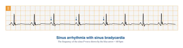
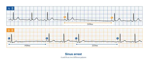
Stock image When sinus arrest occurs, the electrocardiogram will show a long P-P interval, which is not multiples of the basal sinus cycle, including physiological and pathological reasons.
Published: Feb.17, 2024 11:52:04
Author: asia11m
Views: 5
Downloads: 1
File type: image / jpg
File size: 8.96 MB
Orginal size: 10000 x 4418 px
Available sizes:
Level: beginner