Home
Images
Vectors
Videos
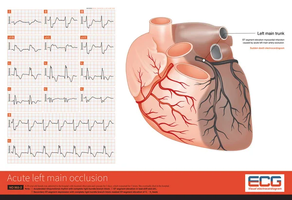
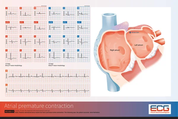
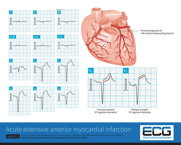
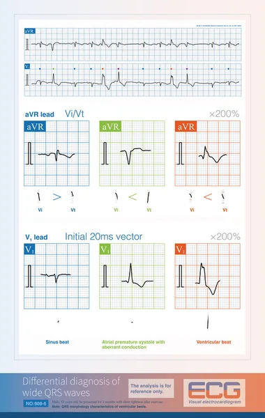
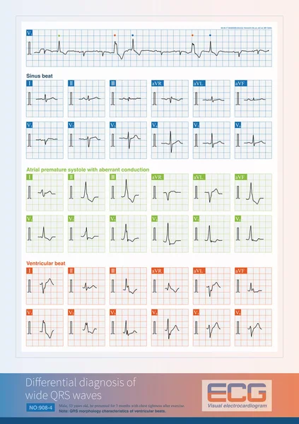
Stock image Atrial focal originating in the left upper pulmonary vein, with an upright P wave in V1 and wide duration, inverted P wave in lead aVL and an upright P wave with notch in inferior leads.
Published:
May.07, 2024 10:55:35
Author:
asia11m
Views:
0
Downloads:
0
File type:
image
/
jpg
File size:
12.65 MB
Orginal size:
10000
x
5632
px
Available sizes:
S
(500 x 282)
M
(1000 x 563)
L
(2000 x 1126)
XL
(10000 x 5632)
EL0
(10000 x 5632)
Level:
beginner
Download Image
Image topics:
Medicine
Wave
Cardiology
Heart
Disease
Beat
Rhythm
Diagnosis
Heartbeat
Cardiac
Ekg
Electrocardiogram
Ecg
Anatomical
Pacemaker
Amaurosis
Ventricle
Arrhythmia
Bradycardia
Heart Disease
Syncope
Cardiac Arrest
Left Ventricle
Superior Vena Cava
Left Atrium
Sudden Death
Inferior Vena Cava
Right Atrium
ECG Monitoring
Coronary Sinus
Bradyarrhythmia
Pulmonary Vein
Sinus Node
P Wave
St Segment
Ecg Education
Qrs Wave
Premature Contraction
Atrial Septum
Pacing Cells
Atrial Premature Contraction
Premature Beats
Ectopic Impulses
Premature Atrial Contractions
Fossa Ovale
Atrial Foci
Ectopic Impulse
Left Upper Pulmonary Vein
Similar stock images
The Coronary Angiography Showed Complete Occlusion Of The Left Main Artery, TIMI 0 And No Stenosis In The Right Coronary Artery With TIMI 3, And A Lateral Circulation To Left Coronary Artery.
10000 × 6859
Male, 65 Years Old, Admitted To Hospital With Chest Pain For 2 Hours. ECG Showed Hyperacute T Waves. Coronary Angiography Showed Occlusion Of The Middle Segment Of Left Anterior Descending Artery.
10000 × 6620
Female, 57 Years Old, Diagnosed With Mitral Stenosis 6 Years Ago. This Electrocardiogram Had An Atrial Premature Contraction, Indicating Its Origin In The Right Superior Pulmonary Vein.
10000 × 6733
Male, 64 Years Old, Admitted With Chest Pain For 4 Hours. The Clinical Diagnosis Was Extensive Anterior And Inferior Myocardial Infarction. The Culprit Vessel Was The Left Anterior Descending Branch.
10000 × 7987
When Atrial Fibrillation Occurs, If The Initial Depolarizations Of A Wide QRS Wave And A Normal QRS Wave Are The Same, It Suggests That The Wide QRS Wave Is Aberrant Conduction.
9000 × 13856
When Analyzing The Nature Of The Wide QRS Complex, Some Indicators Can Be Used In Different ECG Leads, Although The Analysis Does Not Always Achieve 100% Diagnostic Accuracy.
9000 × 14183
A Pre Elderly Woman Suffered From Mitral Stenosis. In 2010, Her Electrocardiogram Showed Sinus Rhythm And Left Atrial Abnormality, And In 2011, She Progressed To Atrial Fibrillation.
10000 × 10258
This ECG Has Two Morphologies Of The Wide QRS Complex And Is Involved In The Differential Diagnosis Of Supraventricular Beats With Aberrant Conduction And Ventricular Beats.
8000 × 11329