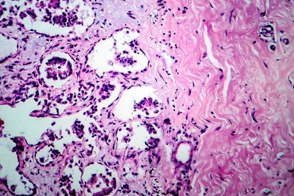
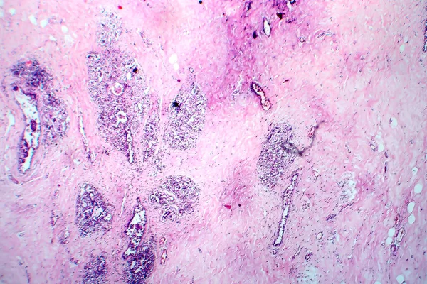
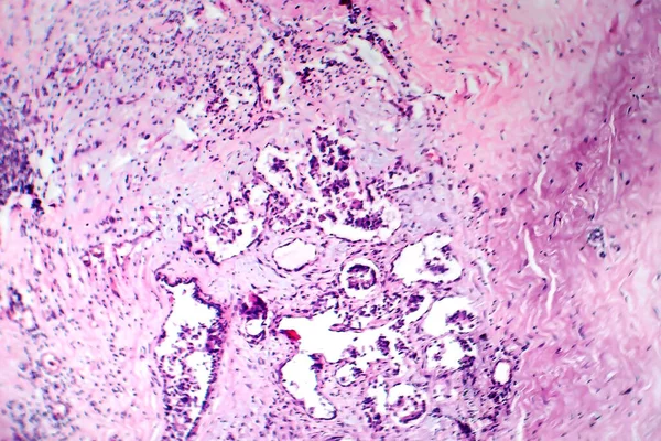
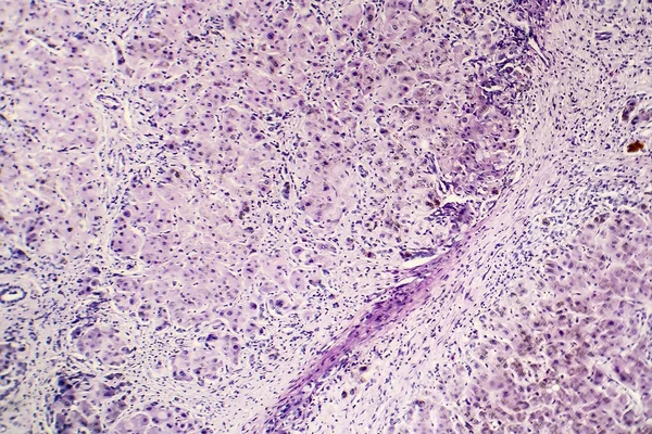
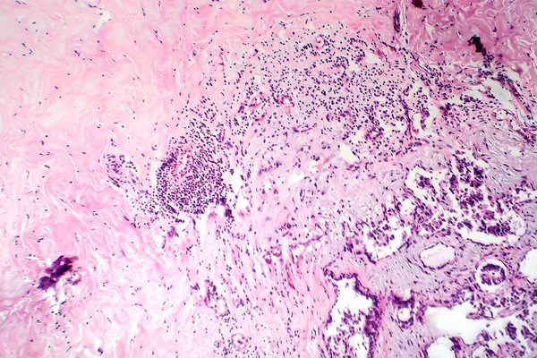
Stock image Breast fibroadenosis, light micrograph, photo under microscope. Common benign hyperplastic process involving breast glands
Published: May.20, 2020 09:18:00
Author: katerynakon
Views: 1
Downloads: 0
File type: image / jpg
File size: 9.51 MB
Orginal size: 4297 x 2865 px
Available sizes:
Level: silver