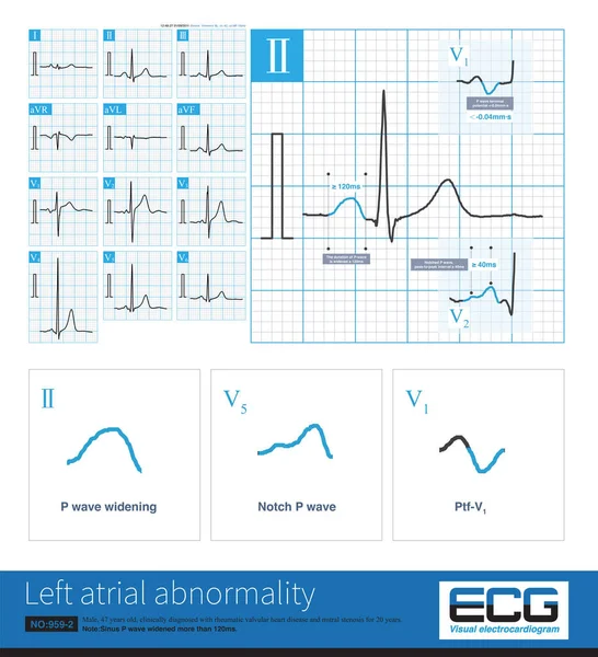
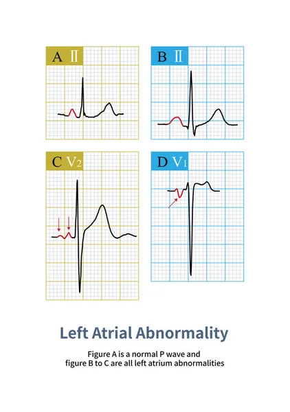
Stock image Clinically, mitral stenosis is a common heart disease affecting the left atrium, and ECG can show notch P wave and P wave widening. Patients will eventually develop atrial fibrillation.

Published: Mar.04, 2024 09:42:52
Author: asia11m
Views: 1
Downloads: 0
File type: image / jpg
File size: 8.95 MB
Orginal size: 10000 x 4684 px
Available sizes:
Level: beginner