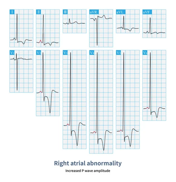
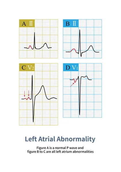
Stock image When the duration and amplitude of sinus P waves increase, it is called a biatrial abnormal ECG change, which usually indicates the presence of lesions in both the left and right atrium.

Published: May.04, 2023 12:22:27
Author: asia11m
Views: 59
Downloads: 0
File type: image / jpg
File size: 14.33 MB
Orginal size: 10000 x 14561 px
Available sizes:
Level: beginner