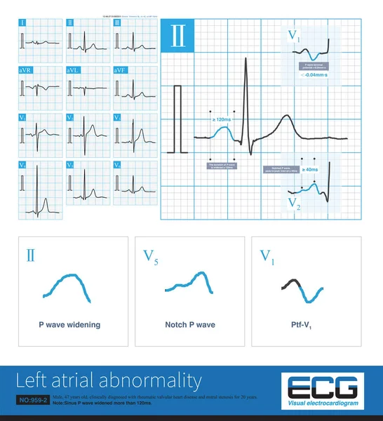
Stock image Female, 52 years old, diagnosed with mitral stenosis 1 years ago. When this ECG was taken, the patient still maintained sinus rhythm.Note that the P wave duration was widened.

Published: May.24, 2023 09:25:05
Author: asia11m
Views: 1
Downloads: 0
File type: image / jpg
File size: 14.36 MB
Orginal size: 10000 x 7768 px
Available sizes:
Level: beginner