photo free
Home Images Vectors Videos

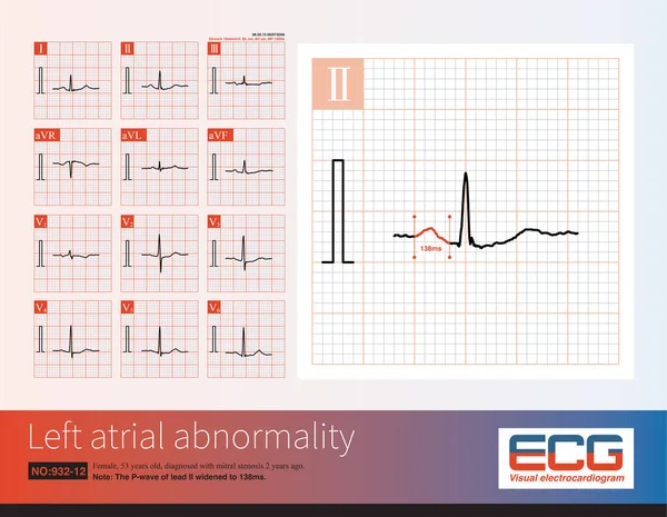
Stock image Female, 56 years old, diagnosed with mitral stenosis 5 years ago. When this ECG was taken, the patient still maintained sinus rhythm.Note that the P wave duration was widened.
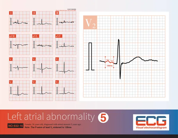
Stock image Female, 56 years old, diagnosed with mitral stenosis 5 years ago. When this ECG was taken, the patient still maintained sinus rhythm.Note that the P wave duration was widened.
Published: May.22, 2023 15:05:43
Author: asia11m
Views: 2
Downloads: 0
File type: image / jpg
File size: 14.37 MB
Orginal size: 10000 x 7768 px
Available sizes:
S
(500 x 388)
M
(1000 x 777)
L
(2000 x 1554)
XL
(10000 x 7768)
EL0
(10000 x 7768)
Level: beginner
Download Image
Image topics:

Similar stock images

stock image Female, 53 years old, diagnosed with mitral stenosis 2 years ago. When this ECG was taken, the patient still maintained sinus rhythm.Note that the P wave duration was widened.

Female, 53 Years Old, Diagnosed With Mitral Stenosis 2 Years Ago. When This ECG Was Taken, The Patient Still Maintained Sinus Rhythm.Note That The P Wave Duration Was Widened.

10000 × 7768
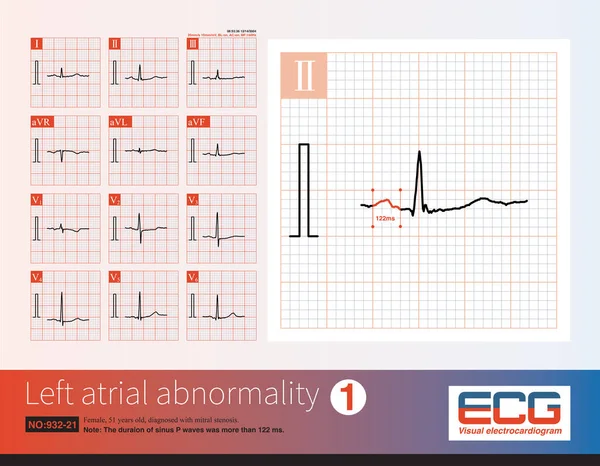
stock image Female, 51 years old, diagnosed with mitral stenosis. When this ECG was taken, the patient still maintained sinus rhythm.Note that the P wave duration was widened.

Female, 51 Years Old, Diagnosed With Mitral Stenosis. When This ECG Was Taken, The Patient Still Maintained Sinus Rhythm.Note That The P Wave Duration Was Widened.

10000 × 7772
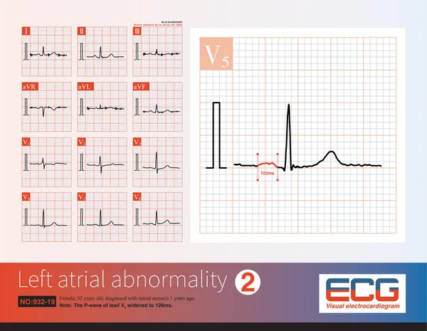
stock image Female, 52 years old, diagnosed with mitral stenosis 1 years ago. When this ECG was taken, the patient still maintained sinus rhythm.Note that the P wave duration was widened.

Female, 52 Years Old, Diagnosed With Mitral Stenosis 1 Years Ago. When This ECG Was Taken, The Patient Still Maintained Sinus Rhythm.Note That The P Wave Duration Was Widened.

10000 × 7768
stock image Female, 57 years old, diagnosed with mitral stenosis 6 years ago. When this ECG was taken, the patient still maintained sinus rhythm and developed atrial fibrillation the following year.

Female, 57 Years Old, Diagnosed With Mitral Stenosis 6 Years Ago. When This ECG Was Taken, The Patient Still Maintained Sinus Rhythm And Developed Atrial Fibrillation The Following Year.

10000 × 4944
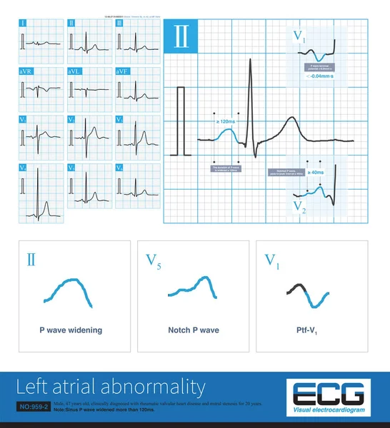
stock image In clinic, mitral stenosis is a common organic heart disease that leads to left atrium abnormality in ECG. The duration of sinus P wave widens by more than 120ms.

In Clinic, Mitral Stenosis Is A Common Organic Heart Disease That Leads To Left Atrium Abnormality In ECG. The Duration Of Sinus P Wave Widens By More Than 120ms.

10000 × 10987
stock image The electrocardiogram characteristics of left atrial abnormality are P wave duration greater than 120ms, P wave notch, peak to peak interval greater than or equal to 40ms, and increased PtfV1.

The Electrocardiogram Characteristics Of Left Atrial Abnormality Are P Wave Duration Greater Than 120ms, P Wave Notch, Peak To Peak Interval Greater Than Or Equal To 40ms, And Increased PtfV1.

10000 × 6534
stock image When the frontal axis is +43 , the maximum QRS depolarization potential is closest to the axis of lead  so the R-wave amplitude of lead  is the highest

When The Frontal Axis Is +43 , The Maximum QRS Depolarization Potential Is Closest To The Axis Of Lead So The R-wave Amplitude Of Lead Is The Highest

10000 × 4944
stock image Female, 57 years old, diagnosed with mitral stenosis 6 years ago. When this ECG was taken, the patient still maintained sinus rhythm and developed atrial fibrillation the following year.

Female, 57 Years Old, Diagnosed With Mitral Stenosis 6 Years Ago. When This ECG Was Taken, The Patient Still Maintained Sinus Rhythm And Developed Atrial Fibrillation The Following Year.

10000 × 4944