Home
Images
Vectors
Videos
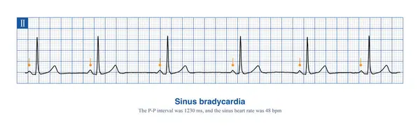
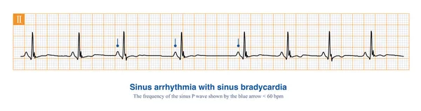
Stock image In clinical practice, sinus arrhythmia often occurs together with sinus bradycardia, most of which are physiological rhythm changes and have no therapeutic significance.
Published:
Feb.17, 2024 06:36:13
Author:
asia11m
Views:
0
Downloads:
0
File type:
image
/
jpg
File size:
5.3 MB
Orginal size:
10000
x
2660
px
Available sizes:
S
(500 x 133)
M
(1000 x 266)
L
(2000 x 532)
XL
(10000 x 2660)
EL0
(10000 x 2660)
Level:
beginner
Download Image
Image topics:
Blue
Background
Horizontal
Graphic
Health
Medical
Care
Cardiology
Heart
Disease
Beat
Rhythm
Diagnosis
Wav
Interval
Duration
Electrode
Cardiogram
Cardiac
Ekg
Electrocardiogram
Atrium
Ecg
Arrhythmia
Electrocardiograph
Bradycardia
Heart Rate
Left Atrium
Right Atrium
Limb Leads
ECG Monitoring
Sinoatrial Node
Sinus Node
Sinus Bradycardia
Sinus Arrhythmia
P Wave
QRS Complex
Cardiovascular Medicine
St Segment
Ecg Education
U Wave
Atrial Muscle
Chest Lead
Cardiac Electrical Activity
Lead Axis
Lead Systems
Pacemaker Cell
Atrial Excitation
Similar stock images
In Clinical Practice, Sinus Arrhythmia Often Occurs Together With Sinus Bradycardia, Most Of Which Are Physiological Rhythm Changes And Have No Therapeutic Significance.
10000 × 2660
When The Frequency Of Sinus Rhythm Is Less Than 60 Beats Per Minute, It Is Called Sinus Bradycardia. Severe Sinus Bradycardia Should Be Suspicious Of Sick Sinus Syndrome.
10000 × 3143
Cardiogram. The Result Of Research CU Is Torn In Half It Is Isolated On A White Background.
4004 × 2558
An 81 Year Old Elderly Male Was Clinically Diagnosed With Coronary Heart Disease And Cerebral Vascular Insufficiency. The Dynamic Electrocardiogram Indicated Sick Sinus Syndrome.
10000 × 1960
When The Frequency Of Sinus Impulses Exceeds 100 Beats Per Minute, Called Sinus Tachycardia, It Is A Common Clinical Arrhythmia. Treatment Focuses On Finding The Cause Of The Increased Heart Rate.
10000 × 7070
A 67-year-old Man Presents With Heart Palpitations, Numbness Of The Lips And Nausea After Consuming Poisonous Shellfish. ECG Showed Sinus Bradycardia.
10000 × 5084
A Is Infantile Type, Short ST Segment, Fast Fusion With T Wave; B Is A Wide Basal T Wave; C Is Normal Type; D Is Late Onset Normal Type, ST Segment Is Prolonged And T Wave Morphology Is Normal.
6500 × 6500
Female, 54 Years Old, Had Recurrent Syncope For 2 Years. The Clinical Diagnosis Was Type 2 Congenital Long QT Syndrome. Her Blood Potassium Is Normal, Do Not Misdiagnose Bimodal T Wave As U Wave.
4000 × 7228