Home
Images
Vectors
Videos
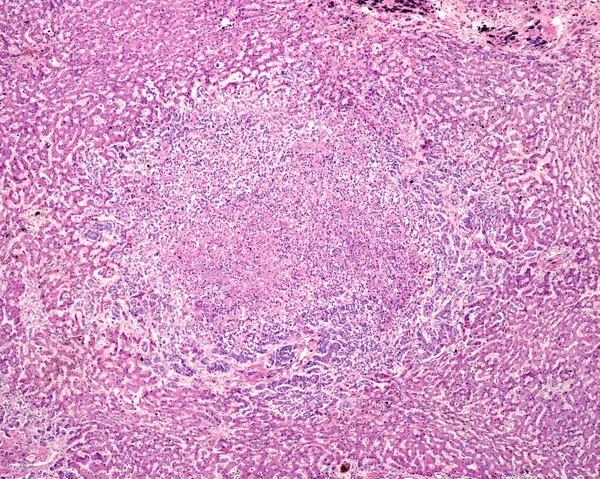
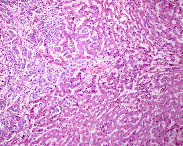
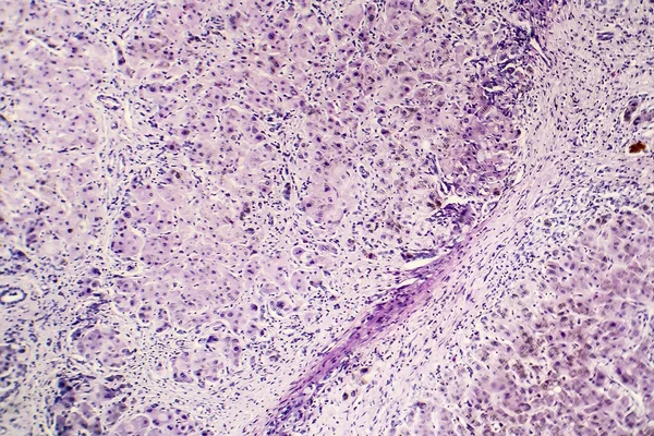
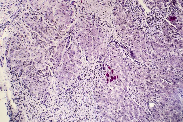
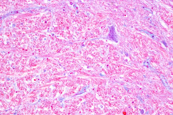
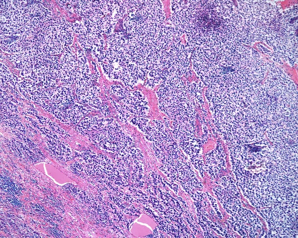
Stock image Light microscope micrograph of a seminoma, a germ cell malignant tumour of the testicle. The micrograph shows sheets of clear cancerous cells with a fibrous stromal network. Darker areas are chronic inflammatory infiltrates.
Published:
Jul.04, 2022 16:30:27
Author:
jlcalvo@ucm.es
Views:
10
Downloads:
0
File type:
image
/
jpg
File size:
16.17 MB
Orginal size:
3840
x
3072
px
Available sizes:
S
(500 x 400)
M
(1000 x 800)
L
(2000 x 1600)
XL
(3840 x 3072)
EL0
(3840 x 3072)
Level:
beginner
Download Image
Image topics:
Human
Medicine
Disease
Cancer
Microscope
Micrograph
Testicle
Microscopy
Tumour
Malignant
Oncology
Oncological
Histology
Light Microscope
Seminoma
Pathology Medical
Similar stock images
Low Magnification Micrograph Of A Seminoma, A Germ Cell Malignant Tumour Of The Testicle. The Micrograph Shows A Lobular Pattern Of Clear Cancerous Cells With A Fibrous Stromal Network.
3840 × 3072
Human Liver. Cholangiocarcinoma. Nodule Of Tumour Cells Showing Central Necrosis And Inflammatory Infiltrates, Surrounded By Normal Hepatocytes.
3840 × 3072
Human Liver. Micronodular Cirrhosis. Regenerating Nodule Of Hepatocytes Showing An Extensive Fatty Change (steatosis), Separated By Fibrous Septa With Chronic Inflammatory Infiltrates.
3840 × 3072
Intrahepatic Cholangiocarcinoma, Ductal Type. Malignant Ductular Structures Formed By Irregular And Complex Glands And Tubules Mixed With A Lymphoplasmacytic Infiltrate.
3840 × 3072
Histopathology Of Postnecrotic Liver Cirrhosis
4147 × 2765
Histopathology Of Postnecrotic Liver Cirrhosis
4260 × 2840
Low Magnification Micrograph Showing A Reticulosarcoma, A Malignant Lymphoma, Infiltrating The Muscular Layer Of The Stomach.
3840 × 3072
Cerebellum, Thalamus, Medulla Oblongata, Spinal Cord And Motor Neuron Human Under The Microscope In Lab.
6000 × 4000