photo free
Home Images Vectors Videos

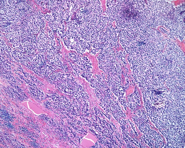
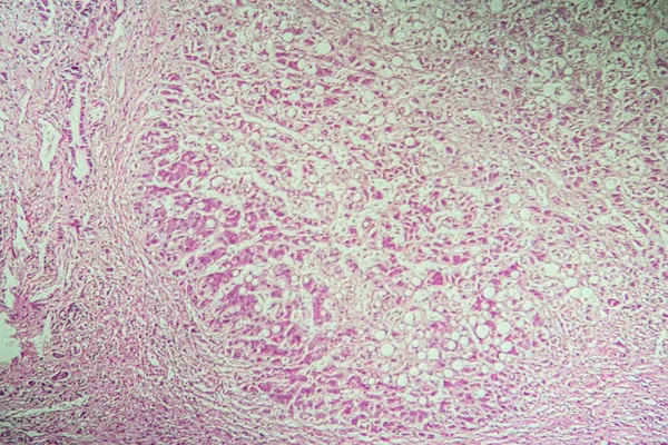
Stock image Low magnification micrograph of a seminoma, a germ cell malignant tumour of the testicle. The micrograph shows a lobular pattern of clear cancerous cells with a fibrous stromal network.

Stock image Low magnification micrograph of a seminoma, a germ cell malignant tumour of the testicle. The micrograph shows a lobular pattern of clear cancerous cells with a fibrous stromal network.
Published: Jul.04, 2022 16:30:27
Author: jlcalvo@ucm.es
Views: 4
Downloads: 0
File type: image / jpg
File size: 16.25 MB
Orginal size: 3840 x 3072 px
Available sizes:
S
(500 x 400)
M
(1000 x 800)
L
(2000 x 1600)
XL
(3840 x 3072)
EL0
(3840 x 3072)
Level: beginner
Download Image
Image topics:

Similar stock images

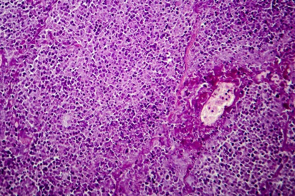
stock image Light microscope micrograph of a seminoma, a germ cell malignant tumour of the testicle. The micrograph shows sheets of clear cancerous cells with a fibrous stromal network. Darker areas are chronic inflammatory infiltrates.

Light Microscope Micrograph Of A Seminoma, A Germ Cell Malignant Tumour Of The Testicle. The Micrograph Shows Sheets Of Clear Cancerous Cells With A Fibrous Stromal Network. Darker Areas Are Chronic Inflammatory Infiltrates.

3840 × 3072
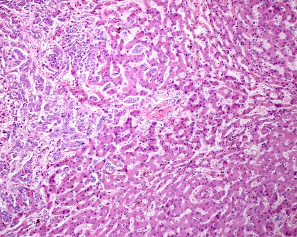
stock image Human liver. Micronodular cirrhosis. Regenerating nodule of hepatocytes showing an extensive fatty change (steatosis), separated by fibrous septa with chronic inflammatory infiltrates.

Human Liver. Micronodular Cirrhosis. Regenerating Nodule Of Hepatocytes Showing An Extensive Fatty Change (steatosis), Separated By Fibrous Septa With Chronic Inflammatory Infiltrates.

3840 × 3072
stock image Intrahepatic cholangiocarcinoma, ductal type. Malignant ductular structures formed by Irregular and complex glands and tubules mixed with a lymphoplasmacytic infiltrate.

Intrahepatic Cholangiocarcinoma, Ductal Type. Malignant Ductular Structures Formed By Irregular And Complex Glands And Tubules Mixed With A Lymphoplasmacytic Infiltrate.

3840 × 3072
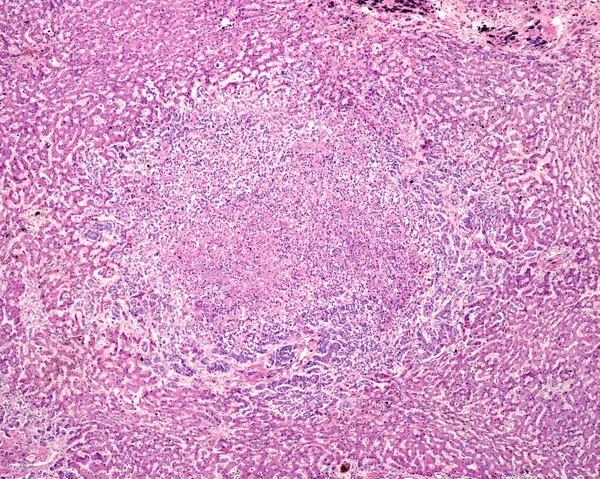
stock image Human liver. Cholangiocarcinoma. Nodule of tumour cells showing central necrosis and inflammatory infiltrates, surrounded by normal hepatocytes.

Human Liver. Cholangiocarcinoma. Nodule Of Tumour Cells Showing Central Necrosis And Inflammatory Infiltrates, Surrounded By Normal Hepatocytes.

3840 × 3072
stock image Human spleen amyloidosis. Amyloidosis is caused by abnormal folding of normal soluble proteins leading to fibril formation. These clumps of protein are called amyloid deposits. The spleen is involved in most cases of systemic amyloidosis. The microgr

Human Spleen Amyloidosis. Amyloidosis Is Caused By Abnormal Folding Of Normal Soluble Proteins Leading To Fibril Formation. These Clumps Of Protein Are Called Amyloid Deposits. The Spleen Is Involved In Most Cases Of Systemic Amyloidosis. The Microgr

3840 × 3072
stock image Caseation of human tuberculosis granuloma, light micrograph

Caseation Of Human Tuberculosis Granuloma, Light Micrograph

4018 × 2679
stock image Histopathology of postnecrotic liver cirrhosis

Histopathology Of Postnecrotic Liver Cirrhosis

4260 × 2840
stock image Liver cirrhosis diseased tissue under the microscope 100x

Liver Cirrhosis Diseased Tissue Under The Microscope 100x

5616 × 3744