
Stock image Right Atrial

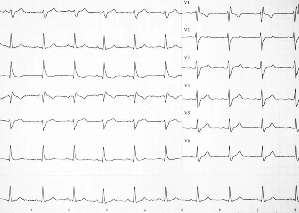
Male, 13 Years Old, Clinically Diagnosed With Secundum Atrial Septal Defect. Note That The QRS Wave In Lead V1 Of The Electrocardiogram Has A QR Shape, Indicating Right Ventricular Hypertrophy.
Image, 7.37MB, 10000 × 7203 jpg
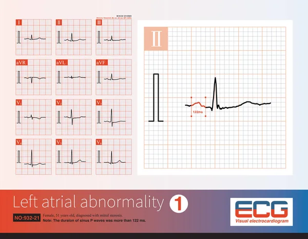
Female, 51 Years Old, Diagnosed With Mitral Stenosis. When This ECG Was Taken, The Patient Still Maintained Sinus Rhythm.Note That The P Wave Duration Was Widened.
Image, 14.21MB, 10000 × 7772 jpg


Cardiac Cycle Diastole And Systole Of Human Heart Anatomy Infographic Diagram With All Stages Of Pumping Filling In Right Left Atrium And Ventricule For Medicine Science Education Medical Healthcare
Vector, 0.66MB, 1913 × 2156 eps
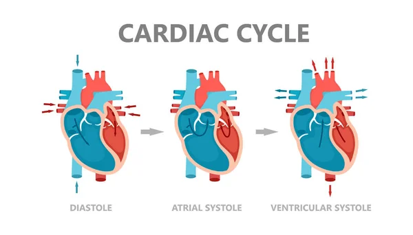
Phases Of The Cardiac Cycle - Diastole, Atrial Systole And Atrial Diastole. Circulation Of Blood Through The Heart. Human Heart Anatomy With Blood Flow.
Vector, 1.18MB, 7000 × 4000 eps
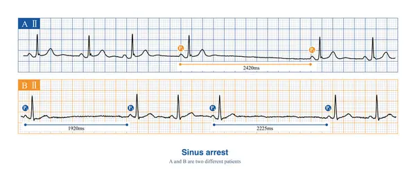
When Sinus Arrest Occurs, The Electrocardiogram Will Show A Long P-P Interval, Which Is Not Multiples Of The Basal Sinus Cycle, Including Physiological And Pathological Reasons.
Image, 8.96MB, 10000 × 4418 jpg

When The Rhythm Of The Atria Originates In The Lower Part Of The Atria, The Whole Atria Are Excited From Inferior To Superior, Producing Negative P Waves In The Inferior Leads.
Image, 11.92MB, 10000 × 8280 jpg

This Photo Shows The Pink Mucinous Matrix Of Atrial Myxoma.Magnify 1000x.
Image, 27.44MB, 4640 × 6303 jpg

This Photo Shows The Pink Mucinous Matrix Of Atrial Myxoma, Myxoma Cells Arranged In Nests And Cords, And Local Bleeding.Magnify 1000x.
Image, 23.48MB, 4640 × 6267 jpg

Phases Of The Cardiac Cycle - Diastole, Atrial Systole And Atrial Diastole. Heart Anatomy Diagram With Blood Flow.
Vector, 0.96MB, 4000 × 4000 eps
Heart Rate On Paper For Recording An Electrocardiogram, Prevention Of Heart Diseases. Electrocardiogram Strips With Cardiac Arrhythmias. Alterations Of Heartbeats Represented On Paper. Copy Space.
Image, 3.11MB, 4675 × 3339 jpg
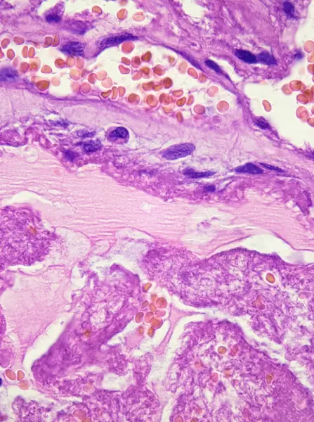
This Photo Shows The Pink Mucinous Matrix Of Atrial Myxoma, Myxoma Cells Arranged In Nests And Cords, And Local Bleeding.Magnify 1000x.
Image, 25.08MB, 4640 × 6226 jpg
Page 1 >> Next