Home
Images
Vectors
Videos
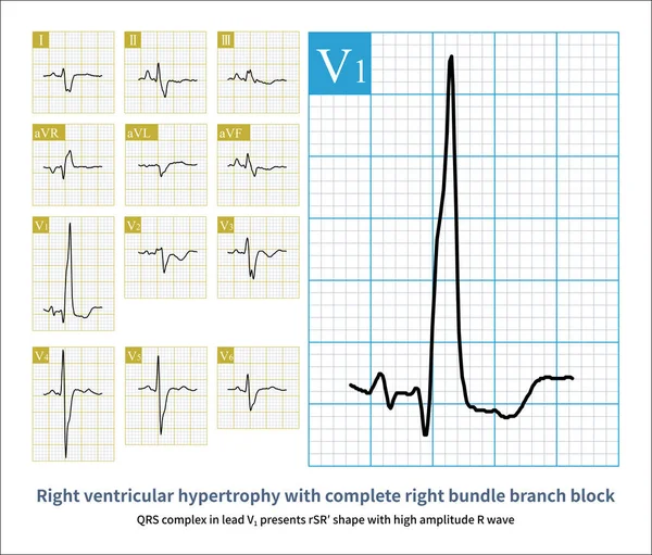
Stock image The standard for diagnosing right atrial abnormality in ECG is that the amplitude of P-wave in limb leadsI is greater than 2.5mm, and the amplitude of upright P-wave in chest leads is 1.5mm.
Published:
Mar.07, 2024 12:50:27
Author:
asia11m
Views:
1
Downloads:
0
File type:
image
/
jpg
File size:
12.42 MB
Orginal size:
10000
x
11279
px
Available sizes:
S
(443 x 500)
M
(887 x 1000)
L
(1773 x 2000)
XL
(10000 x 11279)
EL0
(10000 x 11279)
Level:
beginner
Download Image
Image topics:
Graphic
Illustration
Design
Human
Health
Medicine
Medical
Cardiology
Hospital
Education
Death
Analysis
Graph
Beat
Rhythm
Diagnosis
Heartbeat
Amplitude
Cardiac
Ekg
Electrocardiogram
Atrium
Ecg
Arrhythmia
Lung Disease
Heart Disease
Blood Vessel
Cardiac Arrest
Coronary Heart Disease
Congenital Heart Disease
Electrophysiology
Coronary Artery
Right Ventricle
Electrical Conduction
Pulmonary Hypertension
Left Atrium
Sudden Death
Right Atrium
ECG Monitoring
P Wave
St Segment
Ecg Education
Qrs Wave
Right Atrial Enlargement
Chronic Cor Pulmonale
Pulmonary P Wave
Right Atrial Hypertrophy
Jugular Venous Distension
Gothic P Wave
Similar stock images
Changes Such As Left Atrial Enlargement, Increased Pressure In The Left Atrium, Fibrosis Or Necrosis Of The Left Atrial Myocardium Can Lead To Left Atrial Abnormalities On The ECG.
10000 × 14201
In Clinic, Mitral Stenosis Is A Common Organic Heart Disease That Leads To Left Atrium Abnormality In ECG. The Duration Of Sinus P Wave Widens By More Than 120ms.
10000 × 10987
The ECG Changes Of Left Atrium Abnormality Include Widening Of P Wave Duration, Bimodal P Wave And Increasing Of P Wave Terminal Potential In Lead V1 Lead P Wave.
10000 × 6644
The Electrocardiogram Characteristic Of Right Atrial Abnormality Is An Increase In P Wave Amplitude, With Limb Leads Exceeding 2.5mm And Chest Leads Exceeding 1.5mm.
10000 × 13345
Female, 56 Years Old, Diagnosed With Mitral Stenosis 5 Years Ago. When This ECG Was Taken, The Patient Still Maintained Sinus Rhythm.Note That The P Wave Duration Was Widened.
10000 × 7768
The ECG Changes Of Left Atrium Abnormality Include Widening Of P Wave Duration, Bimodal P Wave And Increasing Of P Wave Terminal Potential In Lead V1 Lead P Wave.
10000 × 8526
QRS Complex In Lead V1 Presents RSR Shape With High Amplitude R Wave.The Electrocardiogram Of A Patient With Atrial Septal Defect Showed Right Ventricular Hypertrophy With Complete RBBB.
10000 × 8521
Male, 34 Years Old, Clinically Diagnosed With Polycythemia Vera. The Electrocardiogram Indicates Atrial Abnormality And Right Ventricular Hypertrophy.
10000 × 6570