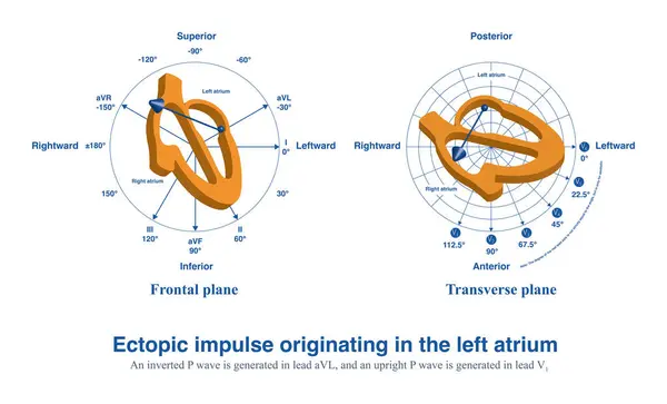
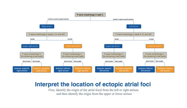
Stock image On a 12 lead electrocardiogram, analyzing the morphology and polarity of ectopic P-waves can determine the approximate anatomical location of ectopic atrial foci.
Published: May.02, 2024 10:48:09
Author: asia11m
Views: 0
Downloads: 0
File type: image / jpg
File size: 6.95 MB
Orginal size: 10000 x 5782 px
Available sizes:
Level: beginner