photo free
Home Images Vectors Videos

Stock image The epicardial electrogram of acute myocardial ischemia was recorded by ligating the left anterior descending branch of the pig. Note T wave inversion during reperfusion.

Stock image The epicardial electrogram of acute myocardial ischemia was recorded by ligating the left anterior descending branch of the pig. Note T wave inversion during reperfusion.
Published: Jun.09, 2022 07:48:26
Author: asia11m
Views: 20
Downloads: 0
File type: image / jpg
File size: 11.65 MB
Orginal size: 10000 x 5216 px
Available sizes:
S
(500 x 261)
M
(1000 x 522)
L
(2000 x 1043)
XL
(10000 x 5216)
EL0
(10000 x 5216)
Level: beginner
Download Image
Image topics:

Similar stock images

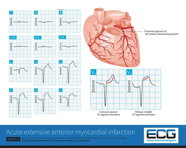
stock image Male, 64 years old, admitted with chest pain for 4 hours. The clinical diagnosis was extensive anterior and inferior myocardial infarction. The culprit vessel was the left anterior descending branch.

Male, 64 Years Old, Admitted With Chest Pain For 4 Hours. The Clinical Diagnosis Was Extensive Anterior And Inferior Myocardial Infarction. The Culprit Vessel Was The Left Anterior Descending Branch.

10000 × 7987
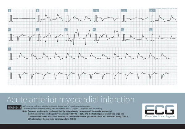
stock image A 69 year old patient with acute myocardial infarction, whose infarction affected the anterior wall, high lateral wall, and inferior wall, ultimately died during hospitalization.

A 69 Year Old Patient With Acute Myocardial Infarction, Whose Infarction Affected The Anterior Wall, High Lateral Wall, And Inferior Wall, Ultimately Died During Hospitalization.

10000 × 6982
stock image Male, 68 years old, chest pain for 7 hours. Coronary angiography suggests occlusion of the distal right coronary artery. The patient was successfully placed with a coronary stent.

Male, 68 Years Old, Chest Pain For 7 Hours. Coronary Angiography Suggests Occlusion Of The Distal Right Coronary Artery. The Patient Was Successfully Placed With A Coronary Stent.

10000 × 8095
stock image Male, 36 years old, without any clinical symptoms. Type B preexcitation was found in ECG, and the deduced accessory pathway was located in the right heart and posterior free wall.

Male, 36 Years Old, Without Any Clinical Symptoms. Type B Preexcitation Was Found In ECG, And The Deduced Accessory Pathway Was Located In The Right Heart And Posterior Free Wall.

10000 × 5878
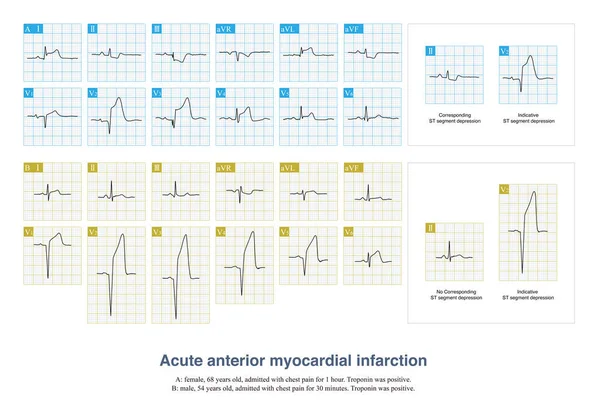
stock image In some patients with acute anterior myocardial infarction, the inferior leads of the electrocardiogram will have ST segment depression, which is called corresponding ST segment depression.

In Some Patients With Acute Anterior Myocardial Infarction, The Inferior Leads Of The Electrocardiogram Will Have ST Segment Depression, Which Is Called Corresponding ST Segment Depression.

10000 × 6954
stock image Normal electrocardiogram of a 34 year old Chinese healthy woman.The rhythm was sinus rhythm without other abnormal ECG changes.

Normal Electrocardiogram Of A 34 Year Old Chinese Healthy Woman.The Rhythm Was Sinus Rhythm Without Other Abnormal ECG Changes.

10000 × 4061
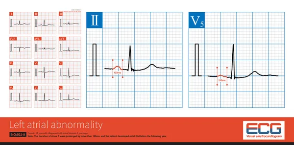
stock image Female, 57 years old, diagnosed with mitral stenosis 6 years ago. When this ECG was taken, the patient still maintained sinus rhythm and developed atrial fibrillation the following year.

Female, 57 Years Old, Diagnosed With Mitral Stenosis 6 Years Ago. When This ECG Was Taken, The Patient Still Maintained Sinus Rhythm And Developed Atrial Fibrillation The Following Year.

10000 × 4944
stock image When the frontal axis is +43 , the maximum QRS depolarization potential is closest to the axis of lead  so the R-wave amplitude of lead  is the highest

When The Frontal Axis Is +43 , The Maximum QRS Depolarization Potential Is Closest To The Axis Of Lead So The R-wave Amplitude Of Lead Is The Highest

10000 × 4944