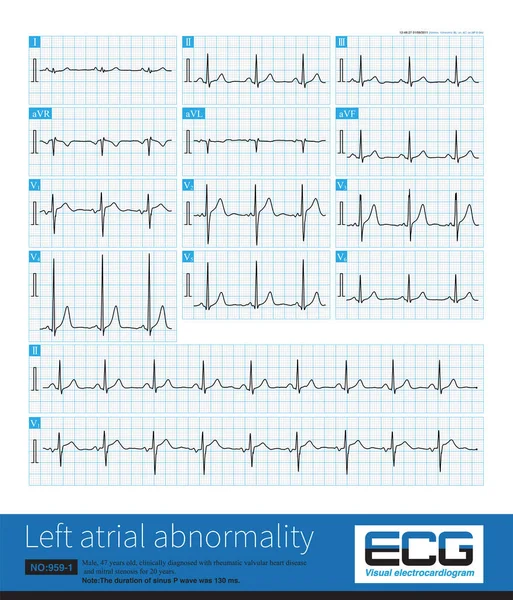
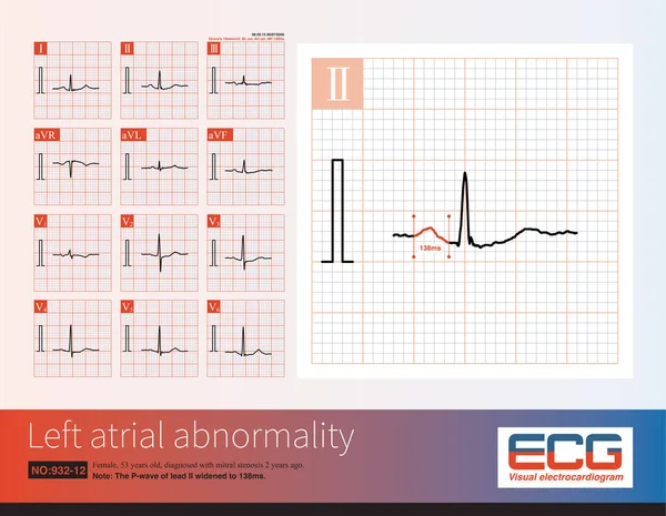
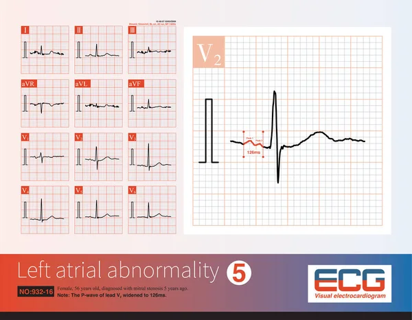
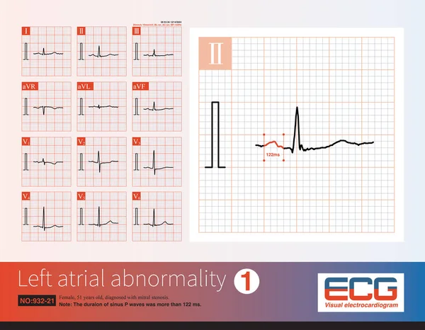
Stock image Female, 57 years old, diagnosed with mitral stenosis 6 years ago. When this ECG was taken, the patient still maintained sinus rhythm and developed atrial fibrillation the following year.

Published: May.08, 2023 08:48:55
Author: asia11m
Views: 2
Downloads: 0
File type: image / jpg
File size: 7.21 MB
Orginal size: 10000 x 4944 px
Available sizes:
Level: beginner