Home
Images
Vectors
Videos
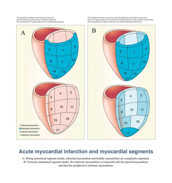
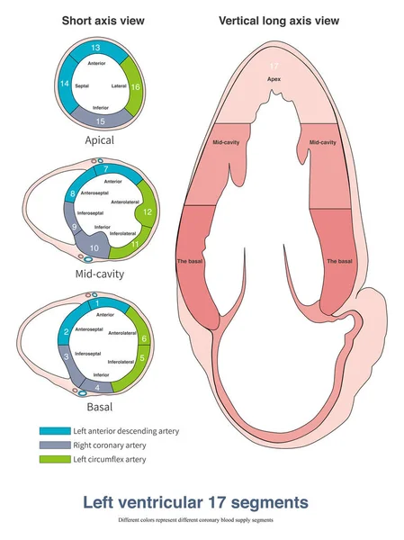
Stock image The left ventricle is divided into 17 segments, and different coronary arteries provide blood supply for different segments.
Published:
Nov.04, 2022 10:56:25
Author:
asia11m
Views:
5
Downloads:
0
File type:
image
/
jpg
File size:
14.03 MB
Orginal size:
10000
x
13358
px
Available sizes:
S
(374 x 500)
M
(749 x 1000)
L
(1497 x 2000)
XL
(10000 x 13358)
EL0
(10000 x 13358)
Level:
beginner
Download Image
Image topics:
White
Blue
Background
Circle
Graphic
Element
Illustration
Design
Shape
Sign
Health
Healthy
Medicine
Healthcare
Medical
Line
Wave
3d
Concept
Icon
Cardiology
Heart
Pulse
Disease
Education
Science
Geometry
Degree
Graph
Beat
Rhythm
Diagnosis
Heartbeat
Cardiogram
Cardiac
Ekg
Heart Rate
Cardiomyopathy
3d Ball
3d Arrow
Sudden Death
Limb Leads
Conduction System
Conduction Disorder
Qrs Wave
Left Axis Deviation
Right Axis Deviation
Frontal Qrs Axis
Wide Qrs Complex
Similar stock images
In The Early Stage Of Acute Transmural Myocardial Ischemia, The Mechanism Of Ventricular Arrhythmia Is Mainly 2 Phase Reentry, Including Reentry Between Ischemic And Non Ischemic Areas And Reentry Across The Ventricular Wall.
10000 × 12923
In Imaging Medicine, Although The Myocardium Is Divided Into Many Segments, In Anatomy, These Myocardium Are Continuous Rather Than Completely Divided.
10000 × 10618
When The Area Or Mass Of Blood Supplied To The Posterior LV By The RCA Exceeds That Of The RV, Acute Inferior MI Also Occurs With A Lead II Elevation Amplitude Greater Than That Of Lead III.
9000 × 13076
Ventricular System Anatomy
4584 × 3789
Hypertrophic Cardiomyopathy Illustration. Apical, Septal And Symmetrical Types
7111 × 4000
Human Skull Anterior View
3840 × 2304
During Ventricular Depolarization, Varying Degrees Of Right Ventricular Hypertrophy And Left Ventricular Confrontation Produce Different Forms Of Right Ventricular Hypertrophy ECG.
10000 × 6096
Chart Of VISUAL SYSTEM - Vector Image
5846 × 4134