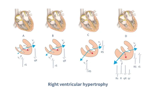
Stock image During ventricular depolarization, varying degrees of right ventricular hypertrophy and left ventricular confrontation produce different forms of right ventricular hypertrophy ECG.
Published: Jun.19, 2023 07:53:06
Author: asia11m
Views: 33
Downloads: 0
File type: image / jpg
File size: 7.51 MB
Orginal size: 10000 x 6096 px
Available sizes:
Level: beginner