
Stock image Chorea page 2
Trembling App Icon. Anxiety. Shaking Body. Worrying And Afraid Person. Chills. Physiological Stress Symptoms.
Vector, 0.75MB, 5000 × 5000 eps
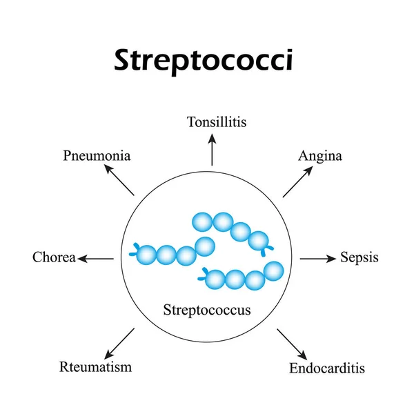
Streptococci. Streptococcal Diseases. Infographics. Vector Illustration
Vector, 3.38MB, 5000 × 5000 eps


Dorsal Striatum Highlighted In Child's Brain And Close-up View Of Its Neurons, 3D Illustration. It Is A Nucleus In The Basal Ganglia, A Component Of The Motor And Reward Systems
Image, 23.52MB, 9339 × 6226 jpg

Destruction Of Neurons Of The Caudate Nucleus, Conceptual 3D Illustration. Caudate Nucleus Belongs To The Brain Basal Ganglia, Its Neurons Are Damaged In Huntingon's Disease And Other Choreas
Image, 16.49MB, 7996 × 5331 jpg

Dorsal Striatum, Caudate Nucleus And Putamen, Highlighted In The Brain Of A Person With Huntington's Disease And Close-up View Of Neuronal Inclusions, Conceptual 3D Illustration
Image, 7.98MB, 6000 × 6000 jpg

Intranuclear Neuronal Inclusions, 3D Illustration. Intranuclear Inclusions In Neurons Are Found In Different Neurodegenerative Diseases, Including Huntingon's Disease, Spinocerebellar Ataxia And Other
Image, 9.43MB, 7200 × 4050 jpg
Trembling Color Icon. Anxiety. Shaking Body. Worrying And Afraid Person. Chills. Physiological Stress Symptoms. Isolated Vector Illustration
Vector, 0.56MB, 5000 × 5000 eps
Tardive Dyskinesia Color Icon. Tremor From Medication. Movement Problem From Neuroleptics. Chorea, Athetosis. Mental Disorder. Neurological Disease From Pills. Isolated Vector Illustration
Vector, 0.63MB, 5000 × 5000 eps
A Person With Huntington's Chorea, A Neurodegenerative Disease Due To Mutation In The HTT Gene, Changes In Brain Basal Ganglia Leading To Random Uncontrollable Movements And Dementia, 3D Illustration
Image, 3.8MB, 3297 × 4946 jpg

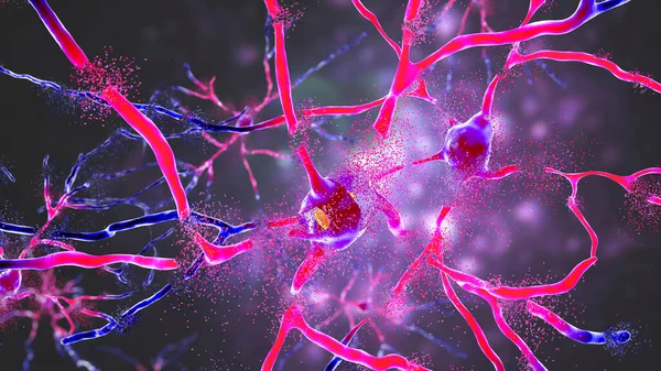
Neuronal Inclusions In Huntington's Disease, 3D Illustration. Inclusions Are Composed Of Mutated Huntingtin Protein, They Are Initially Formed At Axons And Dendrites, Then Migrate To Nuclei Of Neurons
Image, 9.3MB, 7200 × 4050 jpg

Anti-basal Ganglia Antibodies. 3D Conceptual Illustration Showing Molecules Of Immunoglobulins Attacking Dorsal Striatum Highlighted In The Child's Brain. They Are Found In Post-rheumatic Fever Chorea
Image, 25.08MB, 9828 × 6552 jpg
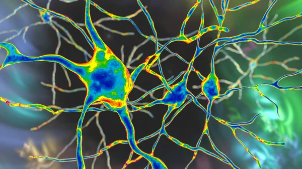
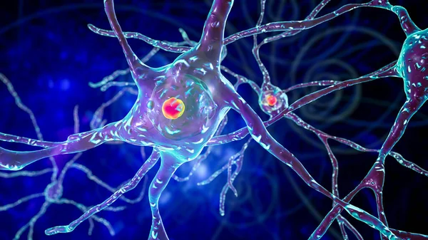
Neurons Of Dorsal Striatum, 3D Illustration. The Dorsal Striatum Is A Nucleus In The Basal Ganglia, Degrading Of Its Neurons Plays A Crucial Role In The Development Of Huntington's Disease
Image, 8.02MB, 7200 × 4050 jpg

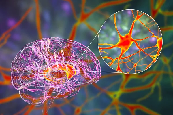
Caudate Nuclei In Human Brain And Its Neurons, 3D Illustration. The Caudate Nucleus Is A Component Of The Basal Ganglia, It Plays Role In Choreas, Neurodegenerative And Other Brain Diseases
Image, 12.8MB, 8157 × 5438 jpg
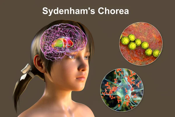
Sydenham's Chorea, An Autoimmune Disease That Results From Streptococcus Infection, Formation Of Anti-neuronal Antibodies Damaging Brain Basal Ganglia That Cause Involuntary Movements, 3D Illustration
Image, 39.11MB, 10609 × 7073 jpg
Neurons Of Dorsal Striatum, 3D Illustration. Dorsal Striatum Is A Nucleus In The Basal Ganglia, Degrading Of Its Neurons Plays Crucial Role In Development Of Huntington's Disease
Image, 12.86MB, 7200 × 4050 jpg
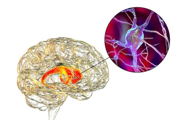
Caudate Nuclei In Human Brain And Its Neurons, 3D Illustration. The Caudate Nucleus Is A Component Of The Basal Ganglia, It Plays Role In Choreas, Neurodegenerative And Other Brain Diseases
Image, 11.51MB, 7996 × 5331 jpg

Neurons Of Dorsal Striatum, 3D Illustration. Dorsal Striatum Is A Nucleus In The Basal Ganglia, Degrading Of Its Neurons Plays Crucial Role In Development Of Huntington's Disease
Image, 8.32MB, 7200 × 4050 jpg

Brain Dorsal Striatum Anatomy, 3D Illustration. The Dorsal Striatum Consists Of The Caudate Nucleus (orange) And The Putamen (blue). Amygdala Is Colored In Red. Front View
Image, 5.28MB, 6000 × 4000 jpg

Intranuclear Neuronal Inclusions, 3D Illustration. Intranuclear Inclusions In Neurons Are Found In Different Neurodegenerative Diseases, Including Huntingon's Disease, Spinocerebellar Ataxia And Other
Image, 8.23MB, 7200 × 4050 jpg

Sydenham's Chorea, An Autoimmune Disease That Results From Streptococcus Infection, Formation Of Anti-neuronal Antibodies Damaging Brain Basal Ganglia That Cause Involuntary Movements, 3D Illustration
Image, 10.26MB, 6947 × 4632 jpg

A Boy With Sydenham's Chorea And Involuntary Movements Of A Hand, 3D Illustration. An Autoimmune Disease After Streptococcus Infection Due To Antibodies Against Cells Of Brain Basal Ganglia And Heart
Image, 8.84MB, 6031 × 4021 jpg
Caudate Nuclei In Human Brain And Its Neurons, 3D Illustration. The Caudate Nucleus Is A Component Of The Basal Ganglia, It Plays Role In Choreas, Neurodegenerative And Other Brain Diseases
Image, 16.77MB, 8157 × 5438 jpg
Trembling Color Icon. Anxiety. Shaking Body. Worrying And Afraid Person. Chills. Physiological Stress Symptoms. Isolated Vector Illustration
Vector, 0.56MB, 5000 × 5000 eps
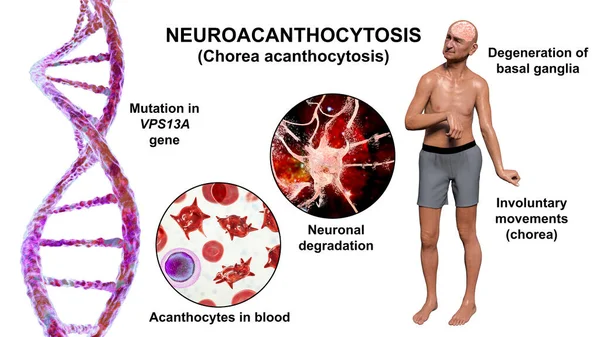
Neuroacanthocytosis, Chorea Acanthocytosis, A Neurodegenerative Disease Due To Mutation In The Gene VPS13A, It Is Marked By Presence Of Acanthocytes In Blood And Choreiform Movements, 3D Illustration
Image, 13.25MB, 9117 × 5128 jpg
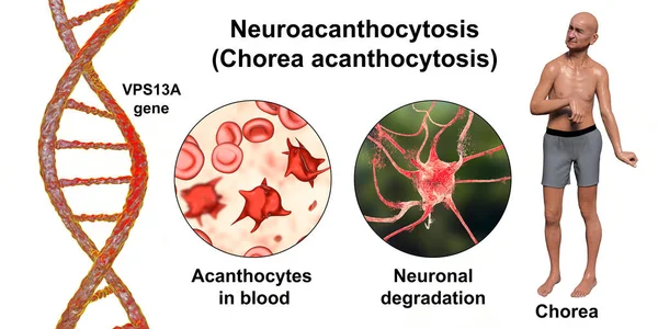
Neuroacanthocytosis, Chorea Acanthocytosis, A Neurodegenerative Disease Due To Mutation In The Gene VPS13A, It Is Marked By Presence Of Acanthocytes In Blood And Choreiform Movements, 3D Illustration
Image, 16.95MB, 10712 × 5356 jpg

Anti-basal Ganglia Antibodies. 3D Conceptual Illustration Showing Molecules Of Immunoglobulins Attacking Dorsal Striatum Highlighted In The Human Brain. They Are Found In Post-rheumatic Fever Chorea
Image, 21.21MB, 10147 × 6765 jpg

Brain In Severe Brain Disease, Dementia, Alzheimer, Chorea Huntington -- 3D Rendering
Image, 19MB, 8000 × 4500 jpg
Previous << Page 2 >> Next