
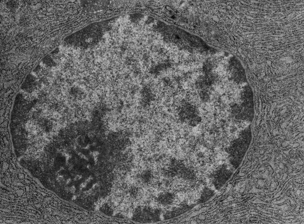
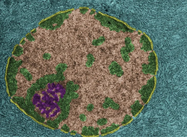
Stock image Serous Cell
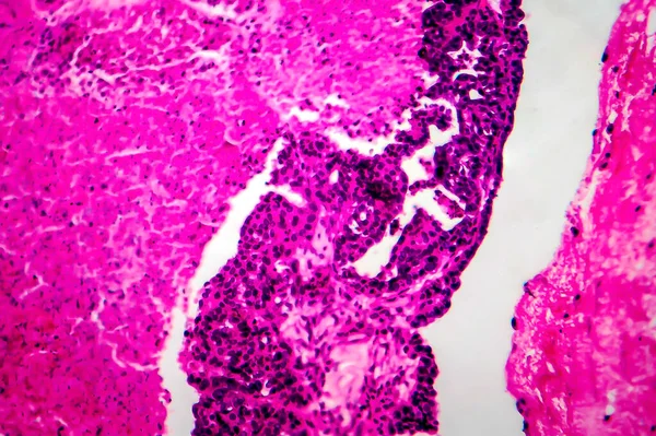
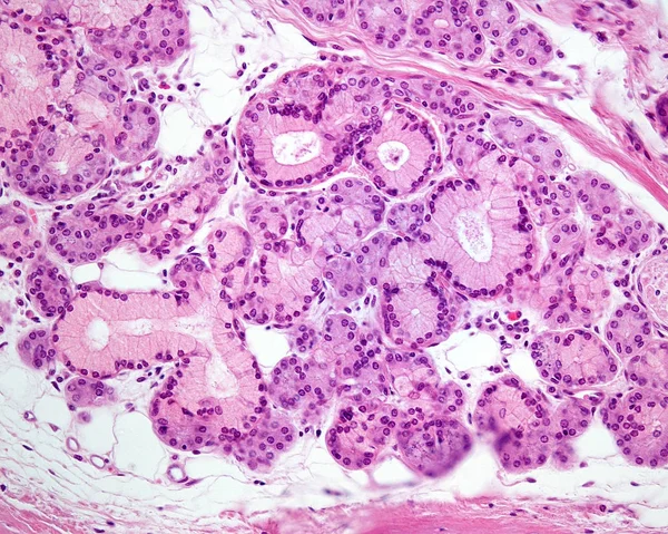
Papillary Serous Ovarian Adenocarcinoma, Cancer Of Ovary, Light Micrograph, Photo Under Microscope
Image, 13.36MB, 4084 × 2722 jpg
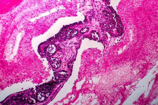
Papillary Serous Ovarian Adenocarcinoma, Cancer Of Ovary, Light Micrograph, Photo Under Microscope
Image, 14.06MB, 4152 × 2768 jpg

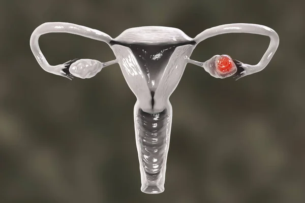
Ovarian Cancer, 3D Illustration Showing Malignant Tumor In The Left Ovary
Image, 6.51MB, 6000 × 4000 jpg
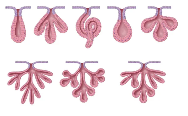
Exocrine Glands Have Two Structural Classifications, Unicellular (one Cell Layer) And Multicellular (many Cell Layers)
Image, 10.06MB, 9449 × 5809 jpg
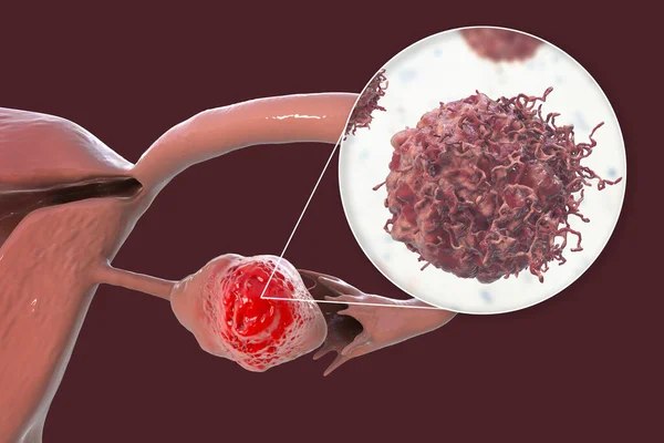
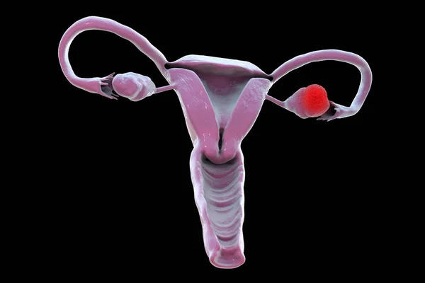
Ovarian Cancer, 3D Illustration Showing Malignant Tumor In The Left Ovary And Close-up View Of Cancer Cells
Image, 8.04MB, 7039 × 4693 jpg
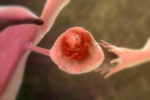
Ovarian Cancer, 3D Illustration Showing Malignant Tumor In The Left Ovary
Image, 10.01MB, 6000 × 4000 jpg
Ovarian Cancer, 3D Illustration Showing Malignant Tumor In The Left Ovary
Image, 2.44MB, 6000 × 4000 jpg
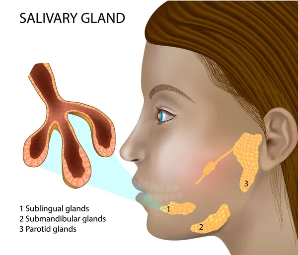
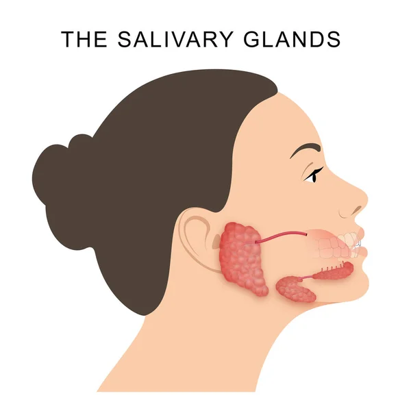
Salivary Gland Structure. Histology Of Salivary Glands. Structure And Cellular Composition Of Mature Salivary Glands.
Vector, 7.76MB, 5001 × 4287 eps
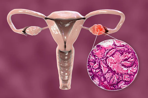
Ovarian Cancer, 3D Illustration Showing Malignant Tumor In The Left Ovary And Light Photomicrograph Showing Histopathology Of Ovarian Cancer
Image, 9.11MB, 6146 × 4097 jpg
Ovarian Cancer, 3D Illustration Showing Malignant Tumor In The Left Ovary
Image, 4.45MB, 6000 × 4000 jpg


This Is A Histological Photograph Of The Human Small Intestine. Magnify 40x.
Image, 9.15MB, 4202 × 4202 jpg

This Is A Pathological Photo Of Human Left Ventricular Hypertrophy, Showing An Increase In Myocardial Diameter And Interstitial Distance.Magnify 40x.
Image, 42.86MB, 8500 × 8500 jpg

This Photo Shows The Pink Mucinous Stroma Of An Atrial Myxoma And The Myxoma Cells Arranged In A Nested And Cord-like Pattern.Magnify 1000x.
Image, 42.93MB, 7000 × 7000 jpg

This Photo Shows The Pink Mucinous Matrix And Nest Like Arrangement Of Mucinous Tumor Cells In Atrial Myxoma.Magnify 1000x.
Image, 42.8MB, 6700 × 6700 jpg

ECF _ Extra Cellular Fluid, Letters And Icons, And Vector Illustration.
Vector, 1.25MB, 3402 × 3402 eps

This Photo Shows Simple Squamous Epithelial Cells On The Surface Of The Human Great Artery, Which Has The Functions Of Exchange And Secretion.
Image, 42.36MB, 8000 × 8000 jpg

This Is A Histological Photograph Of The Human Small Intestine. Magnification 40x
Image, 41.82MB, 8000 × 8000 jpg

This Photo Shows The Pink Mucinous Matrix And Linear Arrangement Of Mucinous Tumor Cells In Atrial Myxoma.Magnify 1000x.
Image, 42.79MB, 6000 × 7851 jpg

This Is A Pathological Photo Of A Human Gout Nodule, Showing The Formation Of Pink Amorphous Eosinophilic Substances By Urate Crystals.Magnify 40x.
Image, 40.54MB, 6000 × 6000 jpg

This Is A Pathological Photo Of Human Left Ventricular Hypertrophy, Showing An Increase In Myocardial Diameter And Interstitial Distance.Magnify 40x.
Image, 40.19MB, 8500 × 8500 jpg


This Is A Pathological Photo Of Human Left Ventricular Hypertrophy, Showing An Increase In Myocardial Diameter And Interstitial Distance.Magnify 40x.
Image, 41.15MB, 8500 × 8500 jpg

This Photo Shows The Pink Mucinous Matrix And Nest Like Arrangement Of Mucinous Tumor Cells In Atrial Myxoma.Magnify 1000x.
Image, 23.51MB, 4640 × 6140 jpg

Gandy-Gamna Bodies Are Pathological Changes Involving Hemosiderosin And Calcium Salt Deposition Produced By Red Blood Cell Decomposition And Fibrous Tissue Encapsulation.
Image, 42.3MB, 6500 × 7799 jpg
Page 1 >> Next