Home
Images
Vectors
Videos
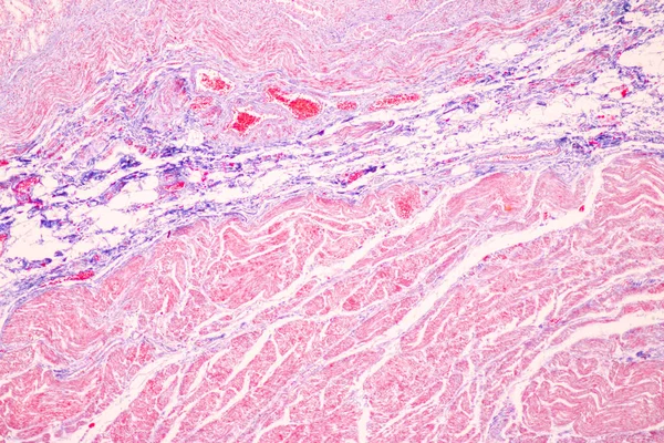
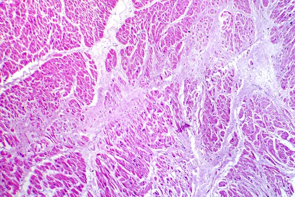
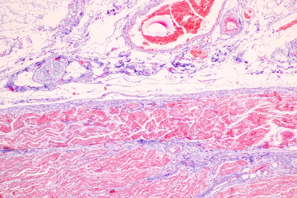
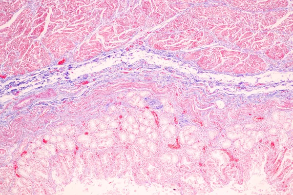
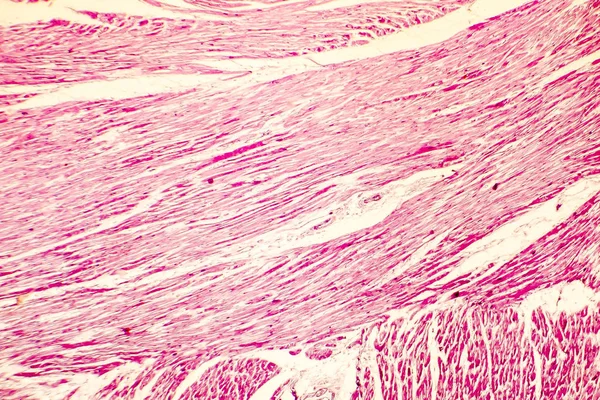
Stock image This is a pathological photo of human left ventricular hypertrophy, showing an increase in myocardial diameter and interstitial distance.Magnify 40x.
Published:
May.11, 2024 08:12:00
Author:
asia11m
Views:
0
Downloads:
0
File type:
image
/
jpg
File size:
41.15 MB
Orginal size:
8500
x
8500
px
Available sizes:
S
(500 x 500)
M
(1000 x 1000)
L
(2000 x 2000)
XL
(8500 x 8500)
EL0
(8500 x 8500)
Level:
beginner
Download Image
Image topics:
Photography
Human
Light
Brown
Medicine
Medical
Heart
Cell
Disease
Area
Elderly
Congestion
Failure
Nucleus
Anatomy
Acute
Deposit
Cardiac
Bleeding
Organs
Infarction
Pathology
Staining
Serous
Nuclei
Congestive
Atrophy
Histology
Ischemia
Cytoplasm
Human Body
Necrosis
Intracellular
Left Ventricle
Right Ventricle
Eosin
Micrography
Hematoxylin
Histopathology
Left Ventricular Hypertrophy
Myocardial Hypertrophy
Stromal Cells
Cardiac Hypertrophy
Myocardial Cells
Basic Medicine
Hypertrophy Of Myocardium
Pathological Sections
Ventricular Muscle Cells
Parenchymal Cells
Similar stock images
Photomicrograph Of Myocardial Infarction, Showing Damaged Heart Tissue Due To Reduced Blood Supply And Cell Death.
5730 × 3821
Papillary Thyroid Carcinoma, Light Micrograph, Photo Under Microscope. The Most Common Type Of Thyroid Cancer
4126 × 2751
Tissue Of Stomach Human Under The Microscope In Lab.
4254 × 2836
Tissue Of Stomach Human Under The Microscope In Lab.
4254 × 2836
Heart Hypertrophy Photomicrograph
3876 × 2584
Tissue Of Small Intestine (Duodenum) And Vermiform Appendix Human Under The Microscope In Lab.
6000 × 4000
Histology Of Human Tissue, Show Chronic Congestion Of Lung As Seen Under The Microscope, 10x Zoom
3115 × 1612
Tissue Of Stomach Human Under The Microscope In Lab.
4254 × 2836