Home
Images
Vectors
Videos
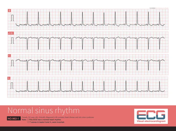
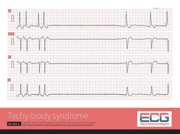
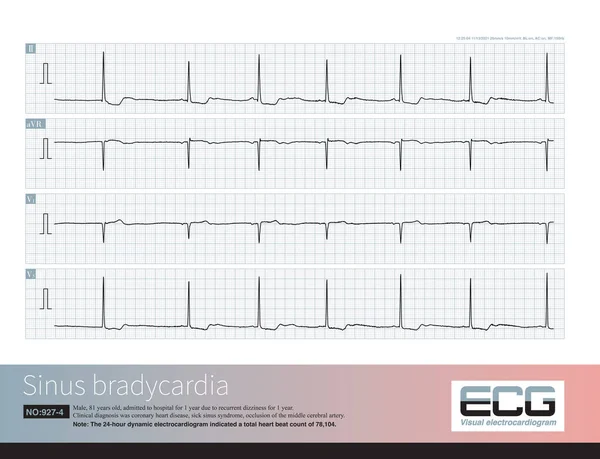
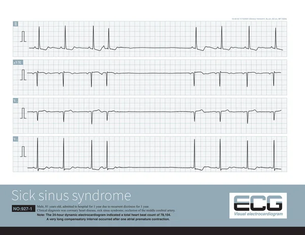
Stock image When sinoatrial node pacemaker cells are dysfunctional, one atrial premature contraction can significantly inhibit the function of sinoatrial node, resulting in a long PP interval.
Published:
May.16, 2023 12:31:00
Author:
asia11m
Views:
8
Downloads:
0
File type:
image
/
jpg
File size:
20.06 MB
Orginal size:
10000
x
7733
px
Available sizes:
S
(500 x 387)
M
(1000 x 773)
L
(2000 x 1547)
XL
(10000 x 7733)
EL0
(10000 x 7733)
Level:
beginner
Download Image
Image topics:
Graphic
Medicine
Medical
Line
Wave
Cardiology
Heart
Monitor
Pulse
Disease
Science
Graph
Rhythm
Escape
Diagnosis
Heartbeat
Cardiac
Ekg
Electrocardiogram
Ecg
Dizziness
Electrocardiograph
Bradycardia
Heart Disease
Syncope
Cardiac Arrest
Coronary Heart Disease
Sudden Death
Sinoatrial Node
Sinus Node
Sinus Arrest
P Wave
Sick Sinus Syndrome
Qt Interval
St Segment
Ecg Education
Left Bundle Branch Block
Death Ecg
Junctional Escape
Pacemaker Cells
Slow Fast Syndrome
Ventricular Escape
Sinus Beat
Escape Rhythm
Atrial Premature Contraction
Slow Arrhythmia
Compensatory Interval
Pairing Interval
Similar stock images
Generally, The Normal Frequency Of Adult Sinus Rhythm Is Between 60 And 100 Bpm, Which Is The Normal Physiological Pacing Frequency Of Sinus Node Pacemaker Cells.
10000 × 7446
After Termination Of Atrial Tachycardia, There Is A Prolonged Sinus Arrest, Followed By A Slow Ventricular Escape, And Finally A Sinus Beat.
10000 × 7447
An 81 Year Old Elderly Male Was Clinically Diagnosed With Coronary Heart Disease And Cerebral Vascular Insufficiency. The Dynamic Electrocardiogram Indicated Sick Sinus Syndrome.
10000 × 10966
A 14-year-old Leukemic Child Had A Sudden Wide QRS Tachycardia With A Frequency Of 167 Bpm, And The Rhythm Was Regular. After Anti-arrhythmia Treatment, The Patient Recovered To Sinus Rhythm.
10000 × 6746
Note That The V3 Lead Of This ECG Shows That The Amplitude Of R Wave Is Greater Than The Amplitude Of S Wave, And There Is Counterclockwise Rotation.
10000 × 11469
An 81 Year Old Elderly Male Was Clinically Diagnosed With Coronary Heart Disease And Cerebral Vascular Insufficiency. The Dynamic Electrocardiogram Indicated Sick Sinus Syndrome.
10000 × 7654
When The Electrical Axis Deviates To The Right, The Maximum QRS Vector Is Located In The Lower Right Quadrant, Which Can Be Interpreted In Combination With The QRS Wave Characteristics Of Limb Leads.
10000 × 7207