
Stock image Ocular Fundus

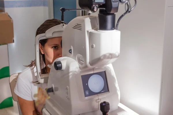
The Patient Is A Doctor With An Ophthalmologist Who Is Watching An Eye Examination Using Modern Technology. Fundus And Optic Nerve Research Concept, Ophthalmoscopy
Image, 4.26MB, 5474 × 3396 jpg
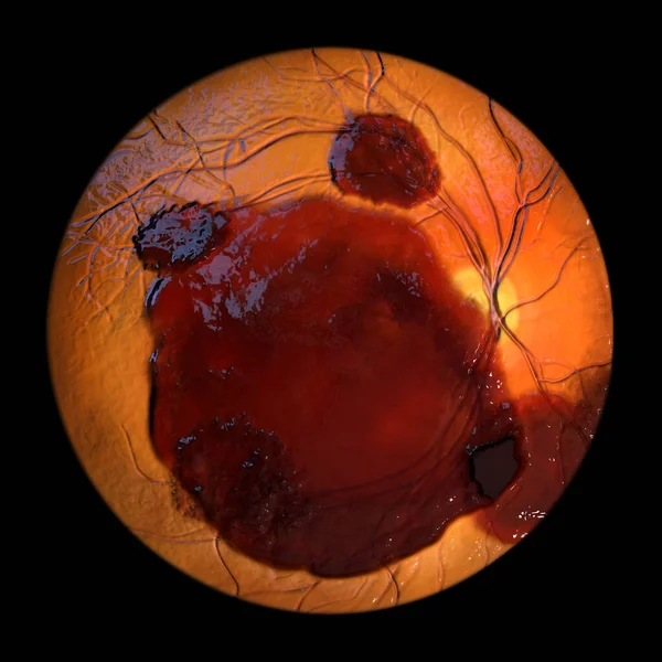
Medical 3D Illustration Of A Subretinal Hemorrhage Observed During Ophthalmoscopy, Revealing A Dark, Irregular Hemorrhage Beneath The Retinal Layers.
Image, 9.37MB, 5352 × 5352 jpg

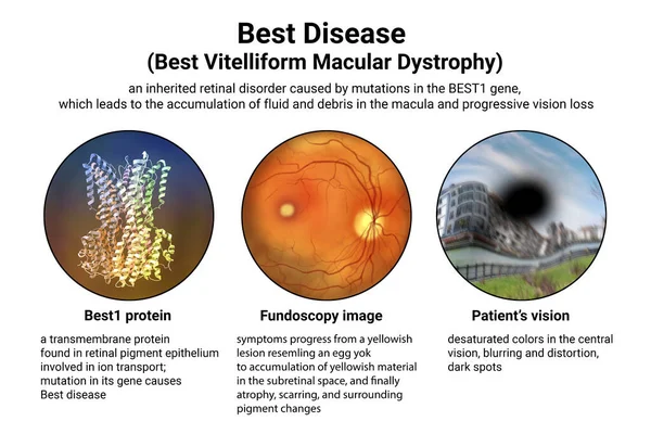
The Difference Between The Vision Of A Normal Eye And An Eye Affected By Best Disease, Illustration Showing Desatured Colors In The Central Vision, Blurring, Distortion, Black Spot
Image, 12.33MB, 9000 × 6000 jpg

Human Eye Anatomy Taking Images With Mydriatic Retinal Cameras. Examination Of The Eye, Diabetic Retinopathy, ARMD
Image, 2.98MB, 3000 × 3186 jpg
Human Eye Anatomy Taking Images With Mydriatic Retinal Cameras. Examination Of The Eye, Diabetic Retinopathy, ARMD
Image, 2.83MB, 3000 × 3186 jpg
Best Disease. Best Vitelliform Macular Dystrophy, 3D Illustration Showing Best1 Protein, Classic Fundoscopic Egg-yolk Lesion On Retina, And Distorted Vision With Black Spot In A Patient
Image, 7.56MB, 9000 × 6000 jpg

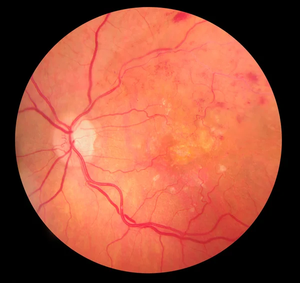
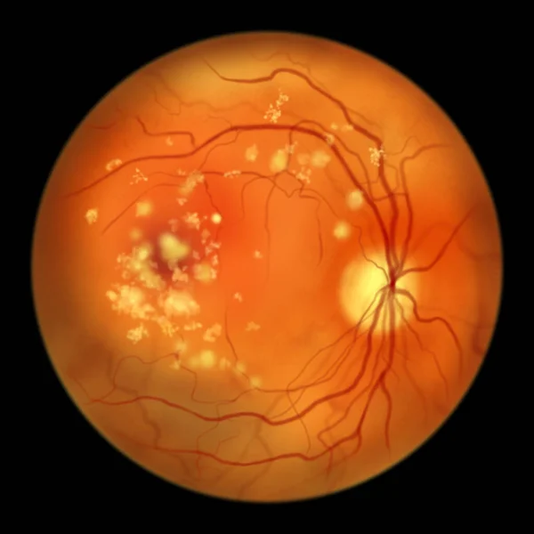
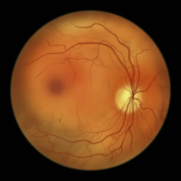
Best Disease. Best Vitelliform Macular Dystrophy, Pseudohypopyon Stage, Layering Of Lipofuscin, Scientific Illustration, Ophthalmoscope View
Image, 2.89MB, 5000 × 5000 jpg
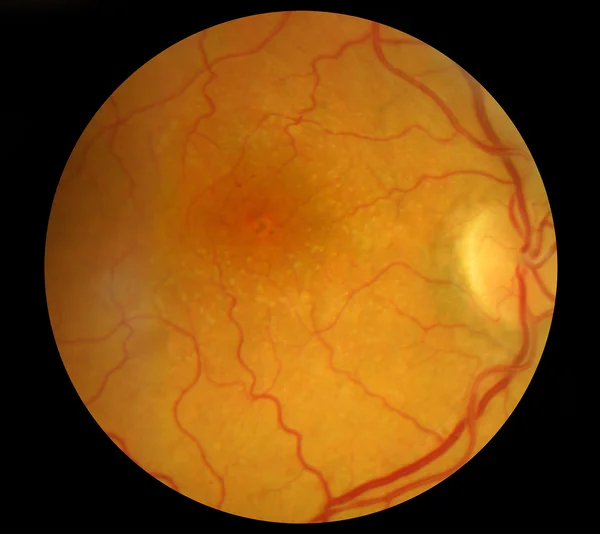
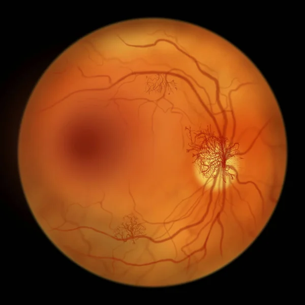
Autosomal Recessive Bestrophinopathy, Ophthalmoscope View, Scientific Illustration Showing Accumulation Of Lipofuscin Deposits Around And Beyond The Macula Leading To Progressive Damage To The Retina
Image, 3MB, 5000 × 5000 jpg

Non-proliferative Diabetic Retinopathy, Illustration Showing Normal Eye Retina And Retina With Hard Exudates, Microaneurysms, Dot Haemorrhages, Flame-shaped And Splinter Retinal Haemorrhages
Image, 7.35MB, 11738 × 6603 jpg

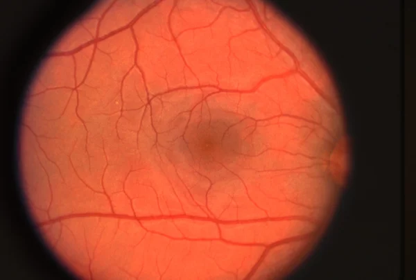
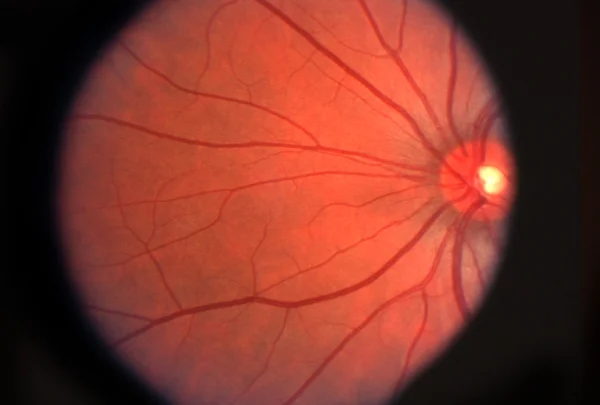
Ophthalmic Image Detailing The Retina And Optic Nerve Inside A Healthy Human Eye. Health Protection Concept
Image, 2.67MB, 3000 × 3186 jpg
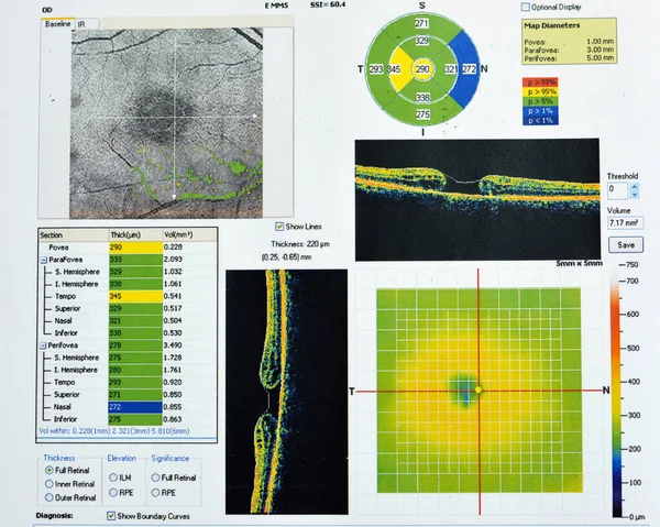
OCT Of The Eye Reveals Faint Epimacular Membrane And Full Thickness Macular Hole Involving The Fovea, Surrounding Diffuse Macular Oedema Showing Few Cystoid Changes For Follow Up, Selective Focus
Image, 11.49MB, 4800 × 3840 jpg

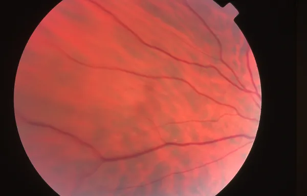
Ophthalmic Image Detailing The Retina And Optic Nerve Inside A Healthy Human Eye. Medicine Concept
Image, 3.76MB, 3000 × 3186 jpg

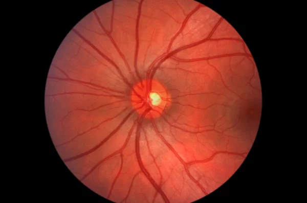
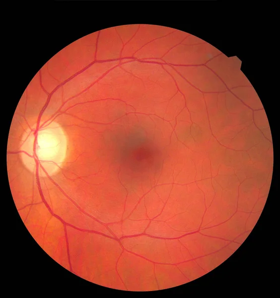
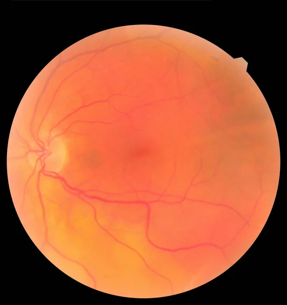
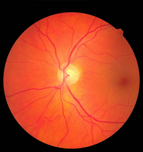
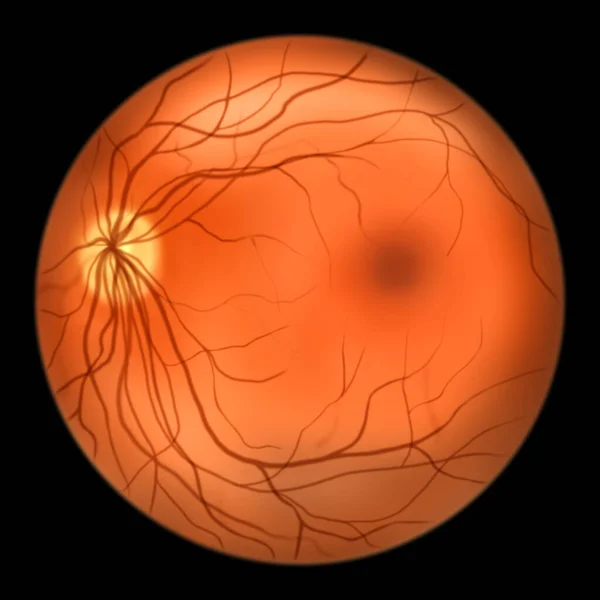
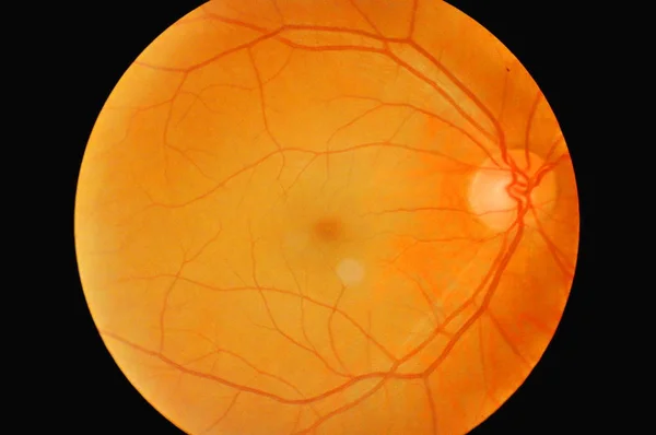
Normal Eye Retina, Ophthalmoscope View, Scientific Illustration Showing Optic Disk, Blood Vessels, Macula And Fovea
Image, 2.74MB, 5000 × 5000 jpg
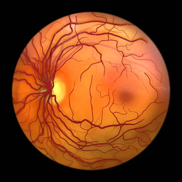
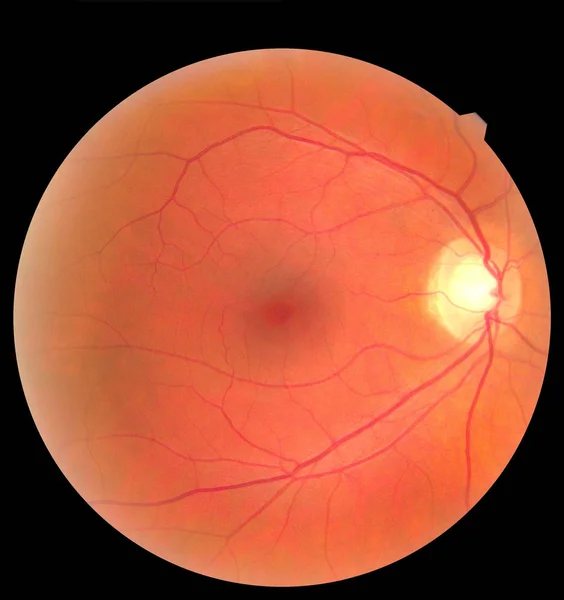
Normal Eye Retina, Ophthalmoscope View, Scientific 3D Illustration Showing Optic Disk, Blood Vessels, Macula And Fovea
Image, 11.53MB, 5000 × 5000 jpg
Ophthalmic Image Detailing The Retina And Optic Nerve Inside A Healthy Human Eye. Health Protection Concept
Image, 2.77MB, 3000 × 3186 jpg

Proliferative Diabetic Retinopathy, Illustration Showing Neovascularization In The Disk And Macula Edema. Abnormal Finding On Fundoscopic Examination Of The Eye Retina In Diabetes Mellitus
Image, 2.85MB, 5000 × 5000 jpg
Proliferative Diabetic Retinopathy, Illustration Showing Neovascularization In The Disk And Other Sites, And Macula Edema. Fundoscopic Examination Of The Eye Retina In Diabetes Mellitus
Image, 2.9MB, 5000 × 5000 jpg
Non-proliferative Diabetic Retinopathy, Illustration Showing IRMAs (intraretinal Microvascular Abnormalities) As Small Vessels With Abnormal Branching Or Dilatation In Ischaemic Areas
Image, 2.68MB, 5000 × 5000 jpg
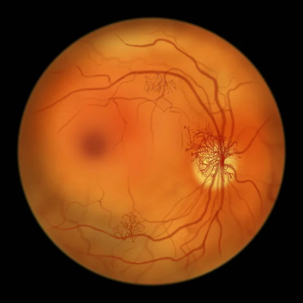
Normal Eye Retina, Ophthalmoscope View, Scientific 3D Illustration Showing Optic Disk, Blood Vessels, Macula And Fovea
Image, 13.02MB, 5352 × 5352 jpg

Diabetic Macular Edema (DME). Diabetic Retinopathy, Illustration Showing Macula Edema, Abnormal Finding On Fundoscopic Examination Of The Eye Retina In Diabetes Mellitus
Image, 2.72MB, 5000 × 5000 jpg

Non-proliferative Diabetic Retinopathy, 3D Illustration Showing Hard Exudates, Microaneurysms, Dot Haemorrhages, Flame-shaped And Splinter Retinal Haemorrhages, Ophthalmoscope View
Image, 13.11MB, 5352 × 5352 jpg
Ophthalmic Image Detailing The Retina And Optic Nerve Inside A Healthy Human Eye. Medicine Concept
Image, 3.41MB, 3000 × 3186 jpg

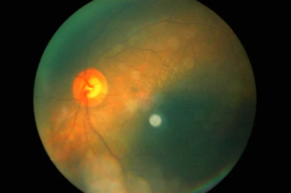
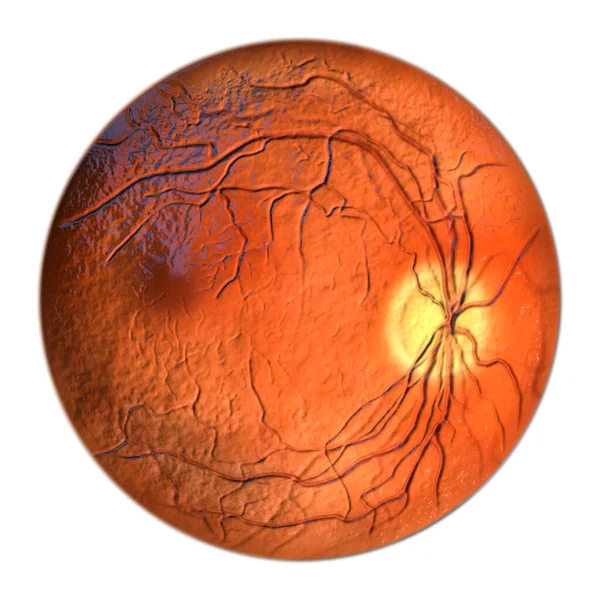
Ocular Toxoplasmosis, A Disease Caused By The Single-celled Protozoan Toxoplasma Gondii. Scientific 3D Illustration Showing Retinal Scar
Image, 8.43MB, 9904 × 6603 jpg

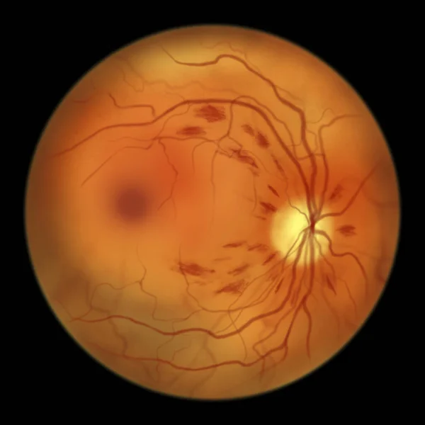
Non-proliferative Diabetic Retinopathy, Illustration Showing Flame-shaped And Splinter Retinal Haemorrhages, Ophthalmoscope View
Image, 2.96MB, 5000 × 5000 jpg
Normal Eye Retina, Ophthalmoscope View, Scientific Illustration Showing Optic Disk, Blood Vessels, Macula And Fovea
Image, 2.88MB, 5000 × 5000 jpg

Non-proliferative Diabetic Retinopathy, 3D Illustration Showing Flame-shaped And Splinter Retinal Haemorrhages, Ophthalmoscope View
Image, 13.41MB, 5352 × 5352 jpg
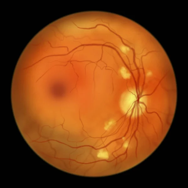
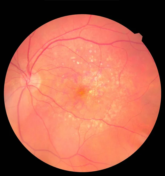
Non-proliferative Diabetic Retinopathy, Illustration Showing Cotton Wool Spots As Fluffy Yellow Patches, Abnormal Finding On Funduscopic Examination Of The Eye Retina In Diabetes Mellitus
Image, 2.7MB, 5000 × 5000 jpg

OCT Of The Eye Reveals Faint Epimacular Membrane And Full Thickness Macular Hole Involving The Fovea, Surrounding Diffuse Macular Oedema Showing Few Cystoid Changes For Follow Up, Selective Focus
Image, 13.79MB, 5541 × 3822 jpg

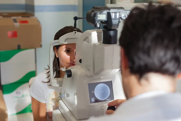
Ophthalmologist Doctor In Exam Optician Laboratory With Female Patient. Eye Care Medical Diagnostic. Eyelid Treatment
Image, 10.02MB, 5624 × 3749 jpg

Proliferative Diabetic Retinopathy, Illustration Showing Neovascularization In The Optic Disk And Other Sites. Fundoscopic Examination Of The Eye Retina In Diabetes Mellitus, Fluorescein Angiography
Image, 6.07MB, 11738 × 6603 jpg
Proliferative Diabetic Retinopathy, Illustration Showing Neovascularization (formation Of New Vessels) In The Optic Disk And Other Sites. Fundoscopic Examination Of The Eye Retina In Diabetes Mellitus
Image, 2.86MB, 5000 × 5000 jpg
Medical Photo Retina Diabetic Retinopathy. Examination Of The Eye, Diabetic Retinopathy, ARMD
Image, 2.56MB, 3000 × 3186 jpg

Non-proliferative Diabetic Retinopathy, Illustration Showing IRMAs (intraretinal Microvascular Abnormalities), Venous Beading, And Microaneurysms
Image, 2.85MB, 5000 × 5000 jpg
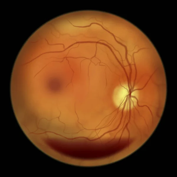
Diabetic Retinopathy, Illustration Shows Preretinal Haemorrhage As Horizontal Blood Level (boat-shaped Haemorrhage), Abnormal Finding On Fundoscopic Examination Of The Eye Retina In Diabetes Mellitus
Image, 2.69MB, 5000 × 5000 jpg

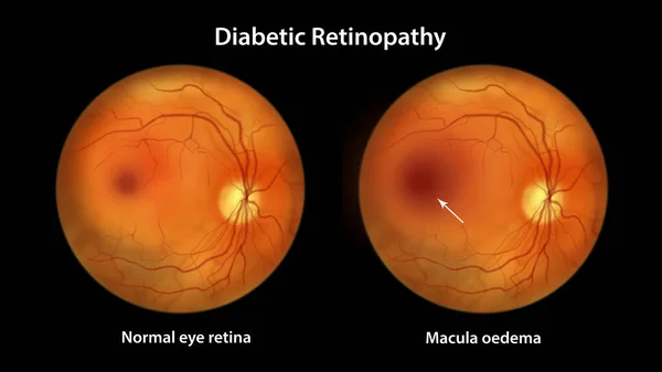
Diabetic Macular Edema (DME), Illustration Showing Normal Eye Retina And Retina With Macula Edema. Fundoscopic Examination Of The Eye Retina In Diabetes Mellitus
Image, 6.24MB, 11738 × 6603 jpg

Non-proliferative Diabetic Retinopathy, 3D Illustration Showing Multiple Microaneurysms On The Eye Retina And Closeup View Of Microaneurysms, Microscopic Buldges In The Artery Walls Filled With Blood
Image, 11.11MB, 10431 × 6954 jpg
Page 1 >> Next