Home
Images
Vectors
Videos
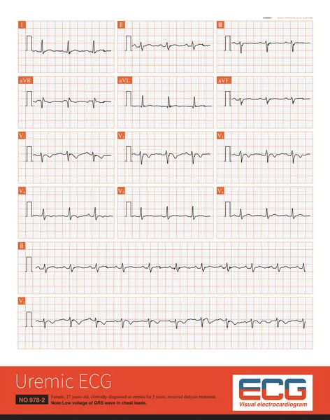
Stock image Female, 27 years old, clinically diagnosed as uremia for 5 years, received dialysis treatment.Chest lead QRS wave showed low voltage.
Published:
May.02, 2022 09:55:27
Author:
asia11m
Views:
9
Downloads:
0
File type:
image
/
jpg
File size:
17.81 MB
Orginal size:
8000
x
10165
px
Available sizes:
S
(394 x 500)
M
(787 x 1000)
L
(1574 x 2000)
XL
(8000 x 10165)
EL0
(8000 x 10165)
Level:
beginner
Download Image
Image topics:
Background
Graphic
Illustration
Design
Human
Medicine
Medical
Line
Cardiology
Heart
Pulse
Information
Disease
Education
Science
Graph
Rhythm
Heartbeat
Potential
Amplitude
Cardiogram
Cardiac
Ekg
Electrocardiogram
Ecg
Myocardium
Dialysis
Depolarization
Cardiomyopathy
Left Ventricle
Right Ventricle
Low Voltage
Uremia
Chronic Renal Insufficiency
P Wave
St Segment
Ecg Education
Qrs Wave
Pericardial Effusion
Chest Leads
St T Change
Limb Lead
Left Axis Deviation
J Point
Initial R Wave
Dialysis Treatment
Chest Lead
Cardiac Electrical Activity
Similar stock images
Normal Electrocardiogram Of A 34 Year Old Chinese Healthy Woman.The Rhythm Was Sinus Rhythm Without Other Abnormal ECG Changes.
4000 × 5237
Cardiac Surgery Is One Of The Common Causes Of Iatrogenic Atrioventricular Block. Once Atrial Fibrillation Exhibits A Slow And Regular Ventricular Rhythm, It Is Important To Be Alert To 3 AVB.
10000 × 11558
Female, 24 Years Old, Healthy. Outpatient Physical Examination ECG Showed Sinus Rhythm, Normal ECG. Note That T-wave Inversion In Lead V1 Is A Normal ECG Phenomenon.
10000 × 11505
When The Frequency Of Sinus Impulses Exceeds 100 Beats Per Minute, Called Sinus Tachycardia, It Is A Common Clinical Arrhythmia. Treatment Focuses On Finding The Cause Of The Increased Heart Rate.
10000 × 7070
The Amplitude Of T Wave In Lead V1 Is Most Commonly Seen In Hypertension And Coronary Heart Disease, Followed By Other Cardiovascular Diseases, And Also Seen In Normal Individuals.
10000 × 6534
Male, 75 Years Old, Clinically Diagnosed As Acute Anterior Septal And High Lateral Myocardial Infarction.The Culprit Vessel Was Located In The LAD Proximal Segment.Prolonged QT Interval With TDP.
10000 × 11911
A 4-year-old Boy With A Clinical Diagnosis Of Long QT Syndrome. No Genetic Testing Was Done During Hospitalization. The Child Died Suddenly During Follow-up.
10000 × 9356