Home
Images
Vectors
Videos
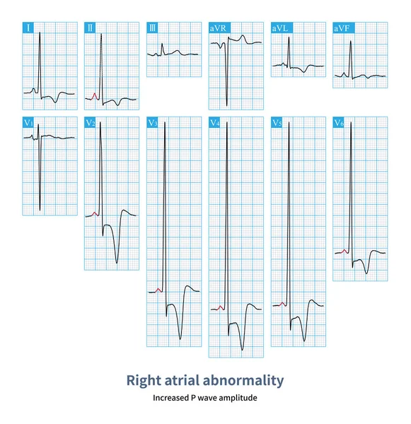
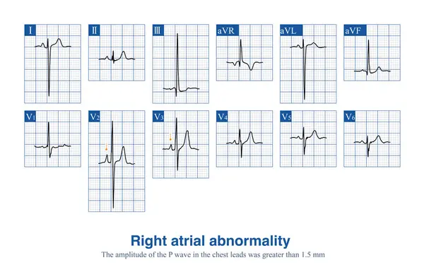
Stock image Male, 10 years old, was clinically diagnosed with tetralogy of Fallot. ECG shows an elevated P wave amplitude in thoracic leads, suggesting right atrial abnormality.
Published:
Apr.08, 2024 09:23:49
Author:
asia11m
Views:
0
Downloads:
0
File type:
image
/
jpg
File size:
11.1 MB
Orginal size:
10000
x
6539
px
Available sizes:
S
(500 x 327)
M
(1000 x 654)
L
(2000 x 1308)
XL
(10000 x 6539)
EL0
(10000 x 6539)
Level:
beginner
Download Image
Image topics:
Design
Human
Health
Medicine
Medical
Cardiology
Hospital
Education
Death
Analysis
Graph
Beat
Rhythm
Diagnosis
Heartbeat
Potential
Amplitude
Cardiac
Ekg
Electrocardiogram
Diaphragm
Atrium
Ecg
Cyanosis
Arrhythmia
Heart Disease
Cardiac Arrest
Congenital Heart Disease
Electrophysiology
Electrical Conduction
Pulmonary Hypertension
Sudden Death
Right Atrium
Ventricular Septal Defect
ECG Monitoring
P Wave
Right Ventricular Hypertrophy
St Segment
Ecg Education
Qrs Wave
Pulmonary Stenosis
Tetralogy Of Fallot
P Waves
Right Atrial Abnormality
Right Atrial Enlargement
Pulmonary P Wave
Jugular Venous Distension
Aortic Straddle
Similar stock images
Female, 46 Years Old, Clinically Diagnosed With Apical Hypertrophic Cardiomyopathy. Pay Attention To The Increased Amplitude Of Sinus P Waves In The Electrocardiogram.
10000 × 10319
Male, 34 Years Old, Clinically Diagnosed With Polycythemia Vera. The Electrocardiogram Indicates Atrial Abnormality And Right Ventricular Hypertrophy.
10000 × 6570
Male, 67 Years Old, With A Clinical Diagnosis Of Chronic Obstructive Pulmonary Disease. ECG Showed Sinus Rhythm And Right Atrial Abnormality.
10000 × 5148
The Standard For Diagnosing Right Atrial Abnormality In ECG Is That The Amplitude Of P-wave In Limb LeadsI Is Greater Than 2.5mm, And The Amplitude Of Upright P-wave In Chest Leads Is 1.5mm.
10000 × 11279
Changes Such As Left Atrial Enlargement, Increased Pressure In The Left Atrium, Fibrosis Or Necrosis Of The Left Atrial Myocardium Can Lead To Left Atrial Abnormalities On The ECG.
10000 × 14201
The ECG Changes Of Left Atrium Abnormality Include Widening Of P Wave Duration, Bimodal P Wave And Increasing Of P Wave Terminal Potential In Lead V1 Lead P Wave.
10000 × 8526
When The Duration And Amplitude Of Sinus P Waves Increase, It Is Called A Biatrial Abnormal ECG Change, Which Usually Indicates The Presence Of Lesions In Both The Left And Right Atrium.
10000 × 14561
Clinically, Mitral Stenosis Is A Common Heart Disease Affecting The Left Atrium, And ECG Can Show Notch P Wave And P Wave Widening. Patients Will Eventually Develop Atrial Fibrillation.
10000 × 4684