Home
Images
Vectors
Videos
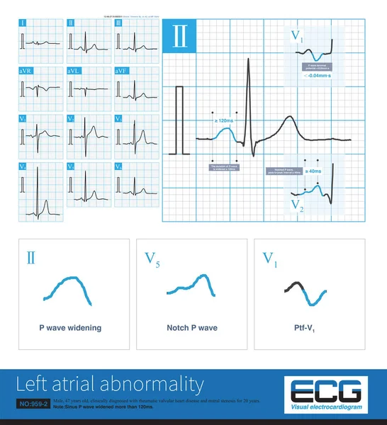
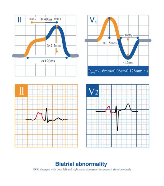
Stock image When the duration of the P wave exceeds 120 ms, the amplitude of the limb leads exceeds 2.5 mm, and the amplitude of the thoracic leads exceeds 1.5 mm, it is interpreted as a biatrial abnormality.
Published:
Apr.12, 2024 12:13:35
Author:
asia11m
Views:
2
Downloads:
0
File type:
image
/
jpg
File size:
12.77 MB
Orginal size:
10000
x
11335
px
Available sizes:
S
(441 x 500)
M
(882 x 1000)
L
(1764 x 2000)
XL
(10000 x 11335)
EL0
(10000 x 11335)
Level:
beginner
Download Image
Image topics:
Design
Human
Health
Medicine
Medical
Cardiology
Hospital
Education
Death
Beat
Rhythm
Diagnosis
Heartbeat
Potential
Amplitude
Cardiac
Ekg
Electrocardiogram
Diaphragm
Atrium
Ecg
Cyanosis
Arrhythmia
Heart Disease
Cardiac Arrest
Congenital Heart Disease
Electrophysiology
Electrical Conduction
Pulmonary Hypertension
Sudden Death
Right Atrium
Ventricular Septal Defect
ECG Monitoring
P Wave
Right Ventricular Hypertrophy
St Segment
Ecg Education
Qrs Wave
Pulmonary Stenosis
Tetralogy Of Fallot
Left Atrial Abnormality
P Waves
Right Atrial Abnormality
Right Atrial Enlargement
Biatrial Abnormality
Jugular Venous Distension
Aortic Straddle
P Wave Amplitude
P Wave Duration
Similar stock images
The Standard For Diagnosing Right Atrial Abnormality In ECG Is That The Amplitude Of P-wave In Limb LeadsI Is Greater Than 2.5mm, And The Amplitude Of Upright P-wave In Chest Leads Is 1.5mm.
10000 × 11279
ECG Paper Is Actually A Coordinate Paper Used For Recording, Consisting Of Small Squares With A Side Length Of 1mm, Representing Time Horizontally And Amplitude Vertically.
10000 × 6473
The ECG Changes Of Left Atrium Abnormality Include Widening Of P Wave Duration, Bimodal P Wave And Increasing Of P Wave Terminal Potential In Lead V1 Lead P Wave.
10000 × 6644
In Clinic, Mitral Stenosis Is A Common Organic Heart Disease That Leads To Left Atrium Abnormality In ECG. The Duration Of Sinus P Wave Widens By More Than 120ms.
10000 × 10987
Male, 36 Years Old, In Good Health, Outpatient Physical Examination ECG. The ECG Is Sinus Rhythm And Normal ECG.
10000 × 7309
Female, 57 Years Old, Diagnosed With Mitral Stenosis 6 Years Ago. When This ECG Was Taken, The Patient Still Maintained Sinus Rhythm And Developed Atrial Fibrillation The Following Year.
10000 × 4944
Female, 57 Years Old, Diagnosed With Mitral Stenosis 6 Years Ago. When This ECG Was Taken, The Patient Still Maintained Sinus Rhythm And Developed Atrial Fibrillation The Following Year.
10000 × 4944
When The Frontal Axis Is +43 , The Maximum QRS Depolarization Potential Is Closest To The Axis Of Lead So The R-wave Amplitude Of Lead Is The Highest
10000 × 4944