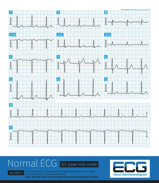
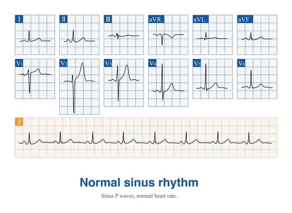
Stock image Male, 36 years old, in good health, outpatient physical examination ECG. The ECG is sinus rhythm and normal ECG.
Published: Jan.03, 2024 20:14:09
Author: asia11m
Views: 2
Downloads: 0
File type: image / jpg
File size: 14.02 MB
Orginal size: 10000 x 7309 px
Available sizes:
Level: beginner