photo free
Home Images Vectors Videos

Stock image When the ST segment is depressed, the amplitude of depression at point J and point J60 can be divided into three types: horizontal (A), downward sloping (B), and upward sloping (C).

Stock image When the ST segment is depressed, the amplitude of depression at point J and point J60 can be divided into three types: horizontal (A), downward sloping (B), and upward sloping (C).
Published: May.02, 2024 10:48:09
Author: asia11m
Views: 5
Downloads: 0
File type: image / jpg
File size: 4.91 MB
Orginal size: 10000 x 4807 px
Available sizes:
S
(500 x 240)
M
(1000 x 481)
L
(2000 x 961)
XL
(10000 x 4807)
EL0
(10000 x 4807)
Level: beginner
Download Image
Image topics:

Similar stock images

stock image According to the offset amplitude of the ST segment at J point and J60 point, ST segment elevation can be divided into three basic types: concave upward, oblique straight and concave downward type.

According To The Offset Amplitude Of The ST Segment At J Point And J60 Point, ST Segment Elevation Can Be Divided Into Three Basic Types: Concave Upward, Oblique Straight And Concave Downward Type.

10000 × 4837
stock image Sometimes, evolution is found on the ECG of a patient with acute myocardial infarction, depending on the time point or lead of the ST-segment elevation.

Sometimes, Evolution Is Found On The ECG Of A Patient With Acute Myocardial Infarction, Depending On The Time Point Or Lead Of The ST-segment Elevation.

10000 × 8921
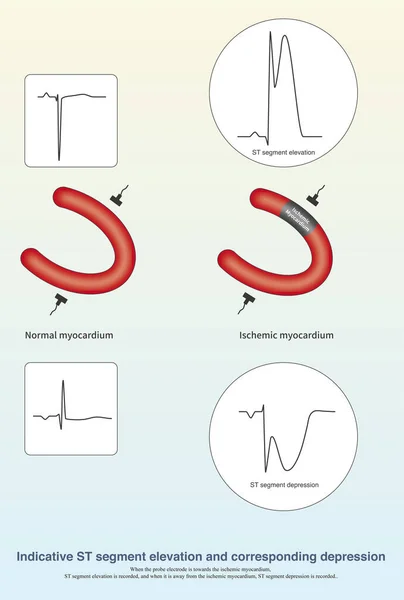
stock image When acute transmural myocardial ischemia occurs, the ST segment of the ischemic myocardium is elevated, and the ST segment of the opposite ischemic myocardium is depressed.

When Acute Transmural Myocardial Ischemia Occurs, The ST Segment Of The Ischemic Myocardium Is Elevated, And The ST Segment Of The Opposite Ischemic Myocardium Is Depressed.

10000 × 14850
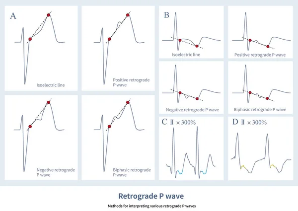
stock image On the ECG, the retrograde P-wave can be a positive wave, a negative wave, a biphasic wave, or an equipotential line wave, which is related to the order of retrograde atrial depolarization.

On The ECG, The Retrograde P-wave Can Be A Positive Wave, A Negative Wave, A Biphasic Wave, Or An Equipotential Line Wave, Which Is Related To The Order Of Retrograde Atrial Depolarization.

10000 × 7093
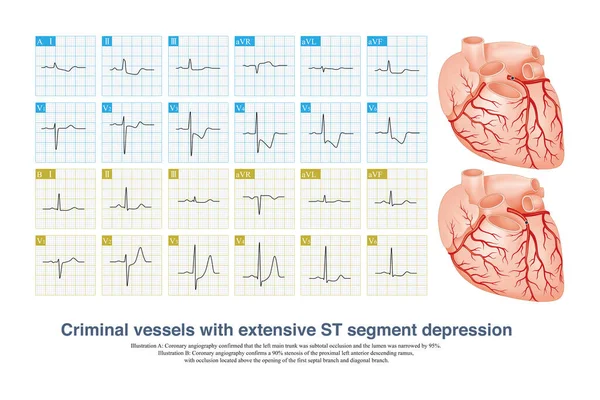
stock image In case of acute coronary syndrome, if the ECG shows extensive ST segment depression, the culprit vessel may be the left main coronary artery or a single coronary artery.

In Case Of Acute Coronary Syndrome, If The ECG Shows Extensive ST Segment Depression, The Culprit Vessel May Be The Left Main Coronary Artery Or A Single Coronary Artery.

10000 × 6672
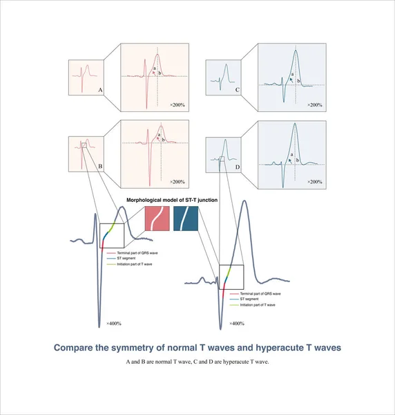
stock image It is worth noting that many electrocardiographic textbooks believe that hyperacute T waves are symmetrical. In fact, they are not symmetrical.

It Is Worth Noting That Many Electrocardiographic Textbooks Believe That Hyperacute T Waves Are Symmetrical. In Fact, They Are Not Symmetrical.

10000 × 10486
stock image Normally, the P wave in lead V1 is either positive or negative biphasic or upright, with the latter noting that a portion of the P wave is actually on the isoelectric line.

Normally, The P Wave In Lead V1 Is Either Positive Or Negative Biphasic Or Upright, With The Latter Noting That A Portion Of The P Wave Is Actually On The Isoelectric Line.

10000 × 11552
stock image Male, 13 years old, clinically diagnosed with secundum atrial septal defect. Note that the QRS wave in lead V1 of the electrocardiogram has a qR shape, indicating right ventricular hypertrophy.

Male, 13 Years Old, Clinically Diagnosed With Secundum Atrial Septal Defect. Note That The QRS Wave In Lead V1 Of The Electrocardiogram Has A QR Shape, Indicating Right Ventricular Hypertrophy.

10000 × 7203