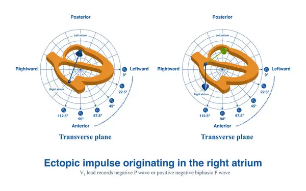
Stock image When ectopic impulses from the anterior wall of the right atrium produce a completely negative P wave in lead V1, the posterior wall ectopic impulse produces a positive and negative biphasic P wave.
Published: Apr.27, 2024 14:15:54
Author: asia11m
Views: 0
Downloads: 0
File type: image / jpg
File size: 8.22 MB
Orginal size: 10000 x 6252 px
Available sizes:
Level: beginner